UniProt data
UniProt name: SIGL7_HUMAN (Q9Y286)
Gene name: SIGLEC7; Synonyms=AIRM1
Organism: Homo sapiens (Human).
Function: Putative adhesion molecule that mediates sialic-acid dependent binding to cells. Preferentially binds to alpha-2,3- and alpha-2,6-linked sialic acid. Also binds disialogangliosides (disialogalactosyl globoside, disialyl lactotetraosylceramide and disialyl GalNAc lactotetraoslylceramide). The sialic acid recognition site may be masked by cis interactions with sialic acids on the same cell surface. In the immune response, may act as an inhibitory receptor upon ligand induced tyrosine phosphorylation by recruiting cytoplasmic phosphatase(s) via their SH2 domain(s) that block signal transduction through dephosphorylation of signaling molecules. Mediates inhibition of natural killer cells cytotoxicity. May play a role in hemopoiesis. Inhibits differentiation of CD34+ cell precursors towards myelomonocytic cell lineage and proliferation of leukemic myeloid cells (in vitro). {ECO:0000269|PubMed:10611343}.
Subcellular location: Membrane; Single-pass type I membrane protein.
Transmembrane domains:
- 354-376, 23 AA, Helical. {ECO:0000255}.
Sequence: 467 AA
CATM prediction
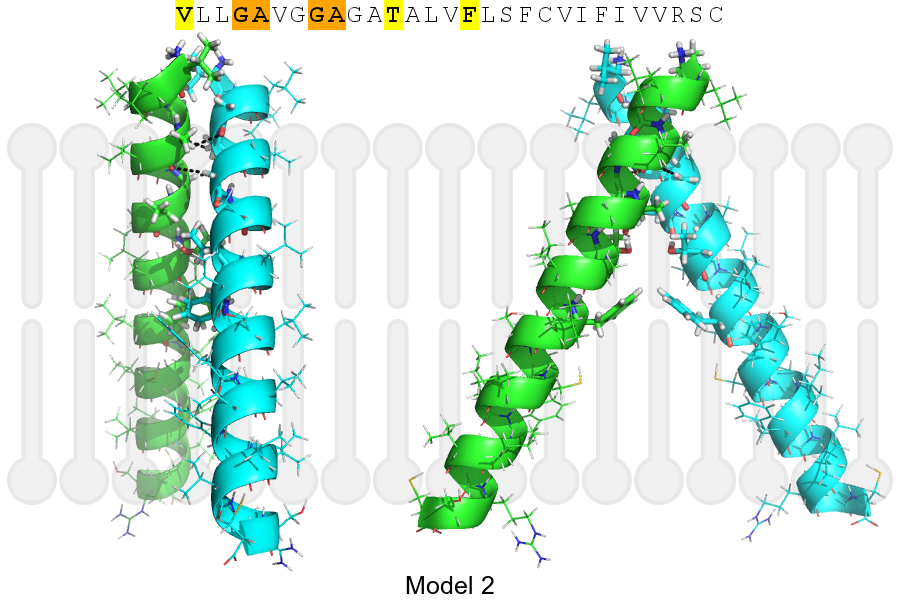
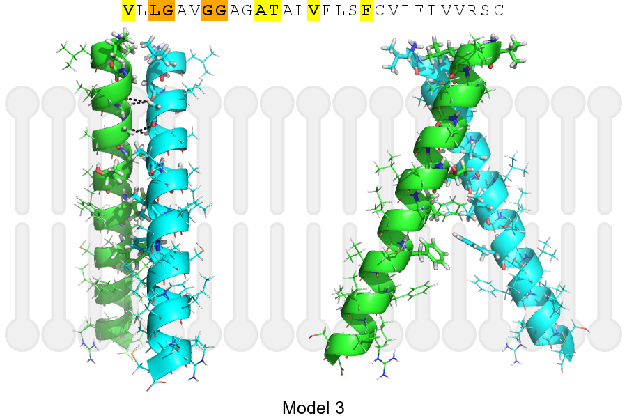
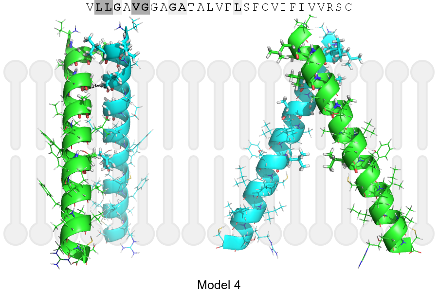
The sequence of SIGL7_HUMAN returned 4 potential models
The positions at the dimer's interface are highlighted in yellow. The position at the crossing point is highlighted in orange.
| # | Sequence and interface | d | θ | Z' | ω' | Score |
|---|---|---|---|---|---|---|
| 360 370 + | + | + |
(Å) | (°) | (Å) | (°) | ||
| Model 1 | VLLGAVGGAGATALVFLSFCVIFIVVRSC | 6.6 | -45.1° | 4.0 | 39.1 | -33.4 |
| Model 2 | VLLGAVGGAGATALVFLSFCVIFIVVRSC | 6.6 | -54.7° | 5.5 | 40.3 | -20.3 |
| Model 3 | VLLGAVGGAGATALVFLSFCVIFIVVRSC | 6.8 | -48.0° | 1.0 | 37.0 | -17.3 |
| Model 4 | VLLGAVGGAGATALVFLSFCVIFIVVRSC | 7.1 | +52.0° | 1.8 | 1.0 | -11.5 |
Click on the model number to visualize its details below.
MODEL 1 DETAILS
Precited structure:
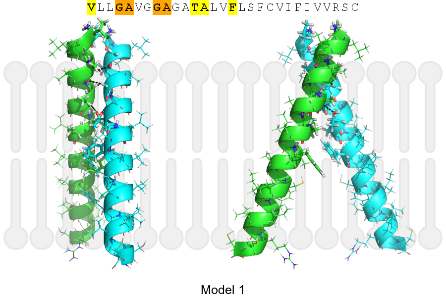
Click on the image to enlarge
Download model 1 structure: PDB, PyMOL session
List of hydrogen bonds:
| Donor | Acceptor | Distance |
|---|---|---|
| B, 355, ALA, HA | A, 354, GLY, O | 2.55Å |
| B, 358, GLY, HA1 | A, 355, ALA, O | 2.73Å |
| B, 359, ALA, HA | A, 362, THR, OG1 | 2.60Å |
| A, 355, ALA, HA | B, 354, GLY, O | 2.55Å |
| A, 358, GLY, HA1 | B, 355, ALA, O | 2.73Å |
| A, 359, ALA, HA | B, 362, THR, OG1 | 2.60Å |