UniProt data
UniProt name: MGT4A_HUMAN (Q9UM21)
Gene name: MGAT4A
Organism: Homo sapiens (Human).
Function: Glycosyltransferase that participates in the transfer of N-acetylglucosamine (GlcNAc) to the core mannose residues of N- linked glycans. Catalyzes the formation of the GlcNAcbeta1-4 branch on the GlcNAcbeta1-2Manalpha1-3 arm of the core structure of N-linked glycans. Essential for the production of tri- and tetra-antennary N-linked sugar chains. Involved in glucose transport by mediating SLC2A2/GLUT2 glycosylation, thereby controlling cell-surface expression of SLC2A2 in pancreatic beta cells.
Subcellular location: Alpha-1,3-mannosyl-glycoprotein 4-beta-N- acetylglucosaminyltransferase A soluble form: Secreted {ECO:0000250}.
Transmembrane domains:
- 5-27, 23 AA, Helical; Signal-anchor for type II membrane protein. {ECO:0000255}.
Sequence: 535 AA
CATM prediction
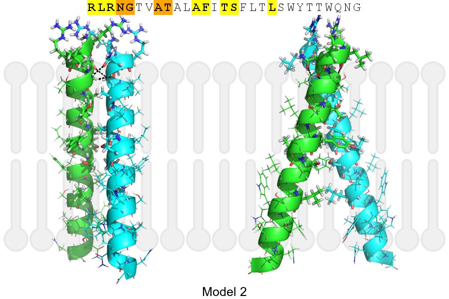
The sequence of MGT4A_HUMAN returned 2 potential models
The positions at the dimer's interface are highlighted in yellow. The position at the crossing point is highlighted in orange.
| # | Sequence and interface | d | θ | Z' | ω' | Score |
|---|---|---|---|---|---|---|
| 10 20 3 + | + | + | |
(Å) | (°) | (Å) | (°) | ||
| Model 1 | RLRNGTVATALAFITSFLTLSWYTTWQNG | 7.4 | -50.0° | 2.9 | 47.0 | -8.0 |
| Model 2 | RLRNGTVATALAFITSFLTLSWYTTWQNG | 7.0 | -36.2° | 3.6 | 36.2 | -4.2 |
Click on the model number to visualize its details below.
MODEL 1 DETAILS
Precited structure:
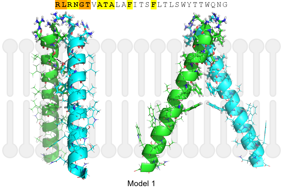
Click on the image to enlarge
Download model 1 structure: PDB, PyMOL session
List of hydrogen bonds:
| Donor | Acceptor | Distance |
|---|---|---|
| B, 7, THR, HA | A, 10, THR, OG1 | 2.76Å |
| A, 7, THR, HA | B, 10, THR, OG1 | 2.76Å |