

UniProt data
UniProt name: HCST_HUMAN (Q9UBK5)
Gene name: HCST; Synonyms=DAP10, KAP10, PIK3AP; ORFNames=UNQ587/PRO1157
Organism: Homo sapiens (Human).
Function: Transmembrane adapter protein which associates with KLRK1 to form an activation receptor KLRK1-HCST in lymphoid and myeloid cells; this receptor plays a major role in triggering cytotoxicity against target cells expressing cell surface ligands such as MHC class I chain-related MICA and MICB, and UL16-binding proteins (ULBPs); these ligands are up-regulated by stress conditions and pathological state such as viral infection and tumor transformation. Functions as docking site for PI3-kinase PIK3R1 and GRB2. Interaction of ULBPs with KLRK1-HCST triggers calcium mobilization and activation of the PIK3R1, MAP2K/ERK, and JAK2/STAT5 signaling pathways. Both PIK3R1 and GRB2 are required for full KLRK1-HCST-mediated activation and ultimate killing of target cells. In NK cells, KLRK1-HCST signaling directly induces cytotoxicity and enhances cytokine production initiated via DAP12/TYROBP-associated receptors. In T-cells, it provides primarily costimulation for TCR-induced signals. KLRK1-HCST receptor plays a role in immune surveillance against tumors and is required for cytolysis of tumors cells; indeed, melanoma cells that do not express KLRK1 ligands escape from immune surveillance mediated by NK cells. {ECO:0000269|PubMed:10426994, ECO:0000269|PubMed:10528161, ECO:0000269|PubMed:11015446, ECO:0000269|PubMed:11777960, ECO:0000269|PubMed:12740575, ECO:0000269|PubMed:12740576, ECO:0000269|PubMed:16002667, ECO:0000269|PubMed:16339517, ECO:0000269|PubMed:16582911, ECO:0000269|PubMed:18097042}.
Subcellular location: Membrane {ECO:0000269|PubMed:10426994}; Single-pass type I membrane protein {ECO:0000269|PubMed:10426994}.
Transmembrane domains:
- 49-69, 21 AA, Helical. {ECO:0000255}.
Sequence: 93 AA
CATM prediction
The sequence of HCST_HUMAN returned 1 potential models
The positions at the dimer's interface are highlighted in yellow. The position at the crossing point is highlighted in orange.
| # | Sequence and interface | d | θ | Z' | ω' | Score |
|---|---|---|---|---|---|---|
| 50 60 70 | + | + | |
(Å) | (°) | (Å) | (°) | ||
| Model 1 | SLALLAGLVAADAVASLLIVGAVFLCA | 7.1 | -40.0° | 4.5 | 44.0 | -24.9 |
| Prolines* | SLPLLAGLVAADAVASLLIVGAVFLCA |
* Note: this sequence contains Pro residues. CATM substituted them with Ala during the modeling. The positions are marked.
Click on the model number to visualize its details below.
MODEL 1 DETAILS
Precited structure:
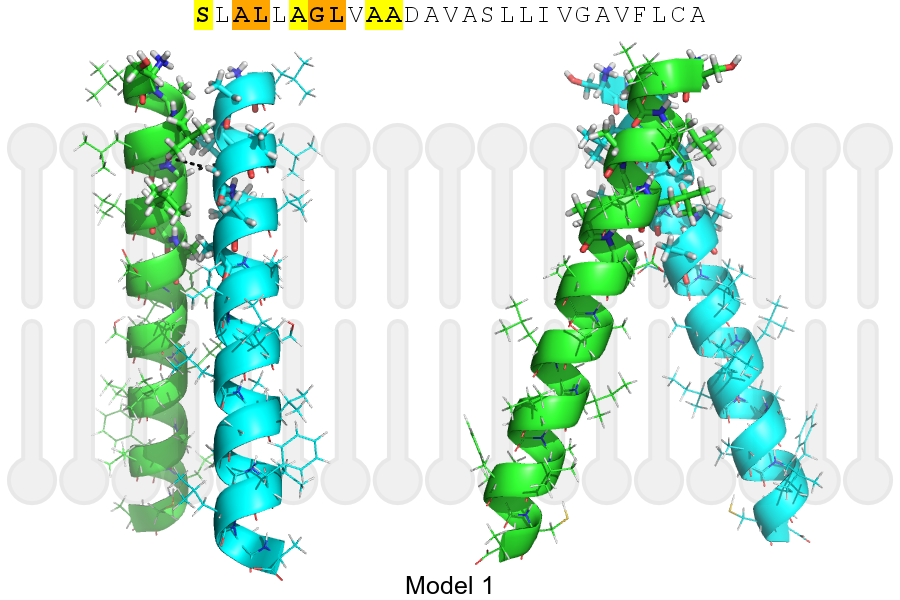
Click on the image to enlarge
Download model 1 structure: PDB, PyMOL session
List of hydrogen bonds:
| Donor | Acceptor | Distance |
|---|---|---|
| B, 52, GLY, HA1 | A, 49, LEU, O | 3.29Å |
| A, 52, GLY, HA1 | B, 49, LEU, O | 3.29Å |