UniProt data
UniProt name: FLRT3_HUMAN (Q9NZU0)
Gene name: FLRT3; Synonyms=KIAA1469; ORFNames=UNQ856/PRO1865
Organism: Homo sapiens (Human).
Function: Functions in cell-cell adhesion, cell migration and axon guidance, exerting an attractive or repulsive role depending on its interaction partners. Plays a role in the spatial organization of brain neurons. Plays a role in vascular development in the retina (By similarity). Plays a role in cell-cell adhesion via its interaction with ADGRL3 and probably also other latrophilins that are expressed at the surface of adjacent cells (PubMed:26235030). Interaction with the intracellular domain of ROBO1 mediates axon attraction towards cells expressing NTN1. Mediates axon growth cone collapse and plays a repulsive role in neuron guidance via its interaction with UNC5B, and possibly also other UNC-5 family members (By similarity). Promotes neurite outgrowth (in vitro) (PubMed:14706654). Mediates cell-cell contacts that promote an increase both in neurite number and in neurite length. Plays a role in the regulation of the density of glutamaergic synapses. Plays a role in fibroblast growth factor-mediated signaling cascades. Required for normal morphogenesis during embryonic development, but not for normal embryonic patterning. Required for normal ventral closure, headfold fusion and definitive endoderm migration during embryonic development. Required for the formation of a normal basement membrane and the maintenance of a normal anterior visceral endoderm during embryonic development (By similarity). {ECO:0000250|UniProtKB:B1H234, ECO:0000250|UniProtKB:Q8BGT1, ECO:0000269|PubMed:14706654, ECO:0000269|PubMed:26235030}.
Subcellular location: Cell membrane {ECO:0000269|PubMed:14706654, ECO:0000269|PubMed:26235030}; Single-pass membrane protein {ECO:0000269|PubMed:14706654, ECO:0000305}. Endoplasmic reticulum membrane {ECO:0000250|UniProtKB:Q8BGT1}. Cell junction, focal adhesion {ECO:0000250|UniProtKB:Q8BGT1}. Secreted {ECO:0000250|UniProtKB:Q8BGT1}. Cell projection, axon {ECO:0000250|UniProtKB:Q8BGT1}. Note=Detected on dendritic punctae that colocalize in part with glutamaergic synapses, but not with GABAergic synapses. Detected on axons and at axon termini. Detected at neuronal growth cones. Proteolytic cleavage in the juxtamembrane region gives rise to a shedded ectodomain. {ECO:0000250|UniProtKB:B1H234, ECO:0000250|UniProtKB:Q8BGT1}.
Transmembrane domains:
- 529-549, 21 AA, Helical. {ECO:0000255}.
Sequence: 649 AA
CATM prediction
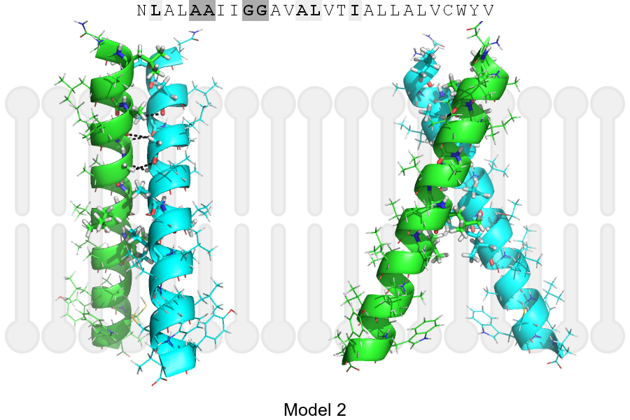
The sequence of FLRT3_HUMAN returned 2 potential models
The positions at the dimer's interface are highlighted in yellow. The position at the crossing point is highlighted in orange.
| # | Sequence and interface | d | θ | Z' | ω' | Score |
|---|---|---|---|---|---|---|
| 530 540 550 | + | + | |
(Å) | (°) | (Å) | (°) | ||
| Model 1 | NLALAAIIGGAVALVTIALLALVCWYV | 6.4 | -48.7° | 1.8 | 45.2 | -35.9 |
| Model 2 | NLALAAIIGGAVALVTIALLALVCWYV | 6.8 | -47.7° | 1.1 | 36.2 | -31.5 |
| Prolines* | NLPLAAIIGGAVALVTIALLALVCWYV |
* Note: this sequence contains Pro residues. CATM substituted them with Ala during the modeling. The positions are marked.
Click on the model number to visualize its details below.
MODEL 1 DETAILS
Precited structure:
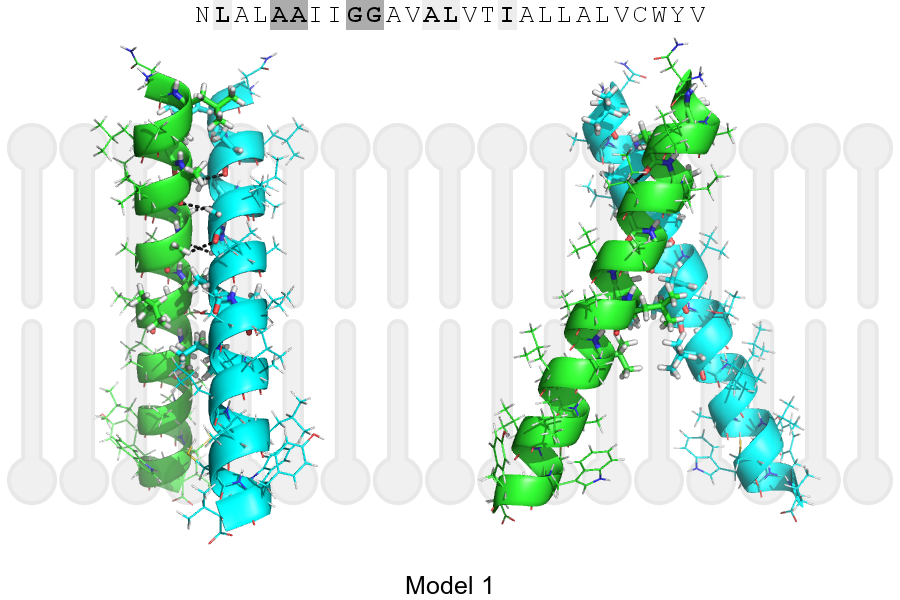
Click on the image to enlarge
Download model 1 structure: PDB, PyMOL session
List of hydrogen bonds:
| Donor | Acceptor | Distance |
|---|---|---|
| B, 531, ALA, HA | A, 530, ALA, O | 2.69Å |
| B, 534, GLY, HA1 | A, 531, ALA, O | 2.36Å |
| B, 535, GLY, HA2 | A, 534, GLY, O | 2.91Å |
| A, 531, ALA, HA | B, 530, ALA, O | 2.69Å |
| A, 534, GLY, HA1 | B, 531, ALA, O | 2.36Å |
| A, 535, GLY, HA2 | B, 534, GLY, O | 2.91Å |