UniProt data
UniProt name: ITM2C_HUMAN (Q9NQX7)
Gene name: ITM2C; Synonyms=BRI3; ORFNames=hucep-14, NPD018, PSEC0047
Organism: Homo sapiens (Human).
Function: Negative regulator of beta amyloid peptide production. May inhibit the processing of APP by blocking its access to alpha- and beta-secretase. Binding to the beta-secretase-cleaved APP C- terminal fragment is negligible, suggesting that ITM2C is a poor gamma-secretase cleavage inhibitor. May play a role in TNF-induced cell death and neuronal differentiation (By similarity). {ECO:0000250, ECO:0000269|PubMed:18452648, ECO:0000269|PubMed:19366692}.
Subcellular location: Lysosome membrane {ECO:0000250}; Single-pass type II membrane protein {ECO:0000250}. Cell membrane {ECO:0000269|PubMed:19114711}; Single-pass type II membrane protein {ECO:0000269|PubMed:19114711}.
Transmembrane domains:
- 55-75, 21 AA, Helical; Signal-anchor for type II membrane protein. {ECO:0000255}.
Sequence: 267 AA
CATM prediction
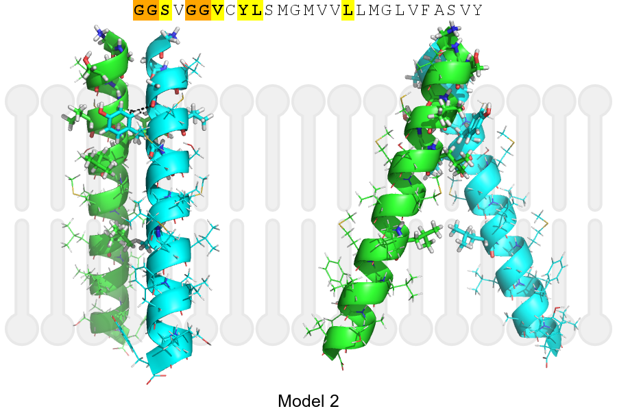
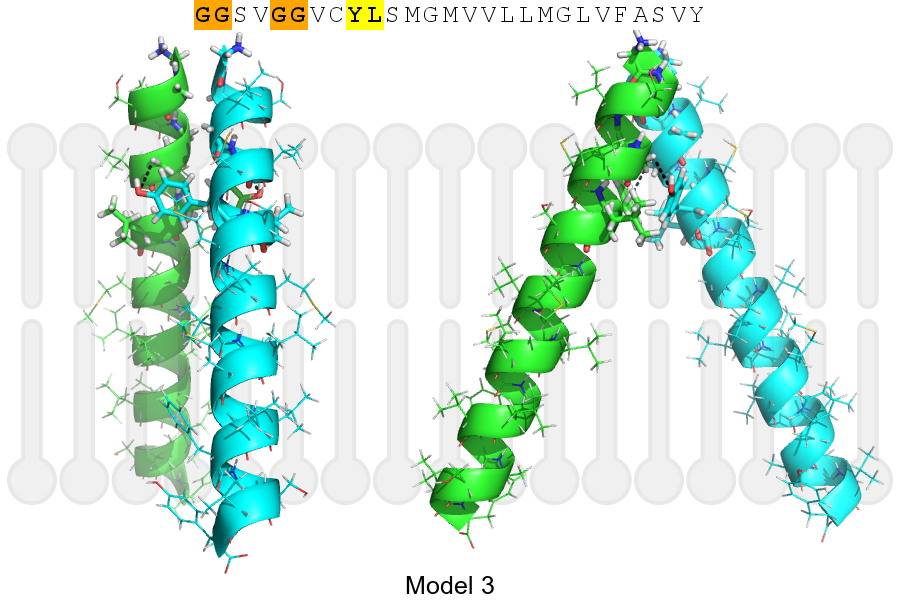
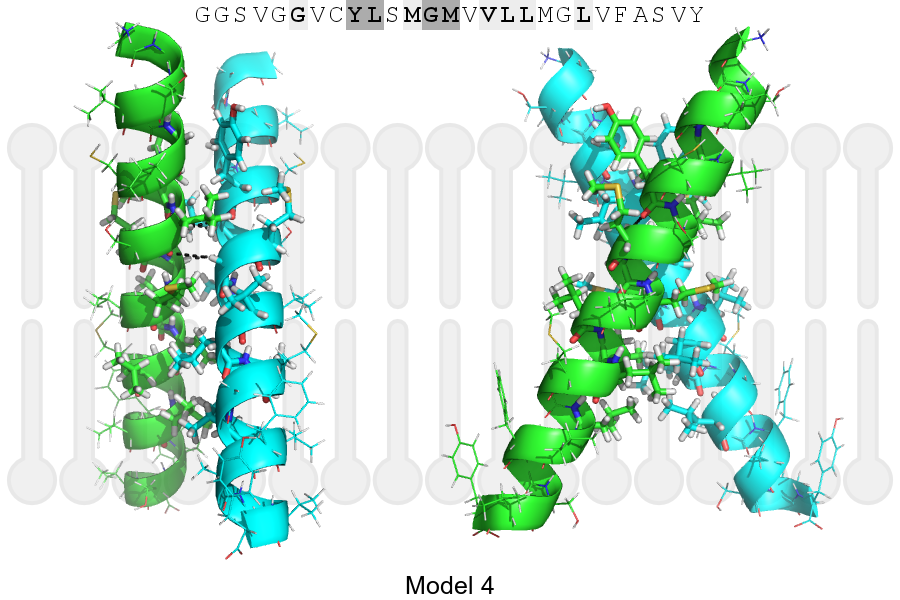
The sequence of ITM2C_HUMAN returned 4 potential models
The positions at the dimer's interface are highlighted in yellow. The position at the crossing point is highlighted in orange.
| # | Sequence and interface | d | θ | Z' | ω' | Score |
|---|---|---|---|---|---|---|
| 60 70 + | + | + |
(Å) | (°) | (Å) | (°) | ||
| Model 1 | GGSVGGVCYLSMGMVVLLMGLVFASVY | 7.4 | -43.0° | 2.4 | 44.0 | -24.2 |
| Model 2 | GGSVGGVCYLSMGMVVLLMGLVFASVY | 7.1 | -42.0° | 1.9 | 33.0 | -7.4 |
| Model 3 | GGSVGGVCYLSMGMVVLLMGLVFASVY | 6.6 | -47.0° | 6.0 | 45.0 | -1.5 |
| Model 4 | GGSVGGVCYLSMGMVVLLMGLVFASVY | 7.2 | -55.2° | 2.4 | 47.8 | -0.4 |
Click on the model number to visualize its details below.
MODEL 1 DETAILS
Precited structure:
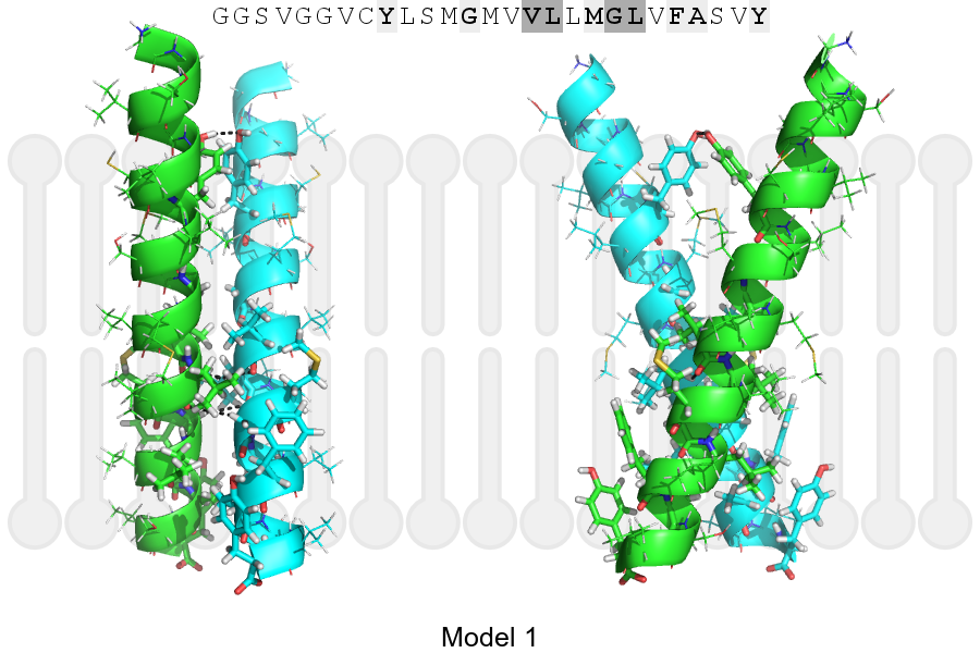
Click on the image to enlarge
Download model 1 structure: PDB, PyMOL session
List of hydrogen bonds:
| Donor | Acceptor | Distance |
|---|---|---|
| B, 68, LEU, HA | A, 67, VAL, O | 3.44Å |
| B, 71, GLY, HA1 | A, 68, LEU, O | 3.36Å |
| A, 60, TYR, HH | B, 60, TYR, OH | 2.11Å |
| A, 68, LEU, HA | B, 67, VAL, O | 3.44Å |
| A, 71, GLY, HA1 | B, 68, LEU, O | 3.36Å |