

UniProt data
UniProt name: CHSTB_HUMAN (Q9NPF2)
Gene name: CHST11
Organism: Homo sapiens (Human).
Function: Catalyzes the transfer of sulfate to position 4 of the N-acetylgalactosamine (GalNAc) residue of chondroitin. Chondroitin sulfate constitutes the predominant proteoglycan present in cartilage and is distributed on the surfaces of many cells and extracellular matrices. Can also sulfate Gal residues in desulfated dermatan sulfate. Preferentially sulfates in GlcA->GalNAc unit than in IdoA->GalNAc unit. Does not form 4, 6- di-O-sulfated GalNAc when chondroitin sulfate C is used as an acceptor.
Subcellular location: Golgi apparatus membrane {ECO:0000250}; Single-pass type II membrane protein {ECO:0000250}.
Transmembrane domains:
- 17-37, 21 AA, Helical; Signal-anchor for type II membrane protein. {ECO:0000255}.
Sequence: 352 AA
CATM prediction
The sequence of CHSTB_HUMAN returned 1 potential models
The positions at the dimer's interface are highlighted in yellow. The position at the crossing point is highlighted in orange.
| # | Sequence and interface | d | θ | Z' | ω' | Score |
|---|---|---|---|---|---|---|
| 20 30 4 + | + | + | |
(Å) | (°) | (Å) | (°) | ||
| Model 1 | ICRMVLATCLGSFILVIFYFQSMLHAV | 7.3 | -54.9° | 4.2 | 37.5 | -11.6 |
| Prolines* | ICRMVLATCLGSFILVIFYFQSMLHPV |
* Note: this sequence contains Pro residues. CATM substituted them with Ala during the modeling. The positions are marked.
Click on the model number to visualize its details below.
MODEL 1 DETAILS
Precited structure:
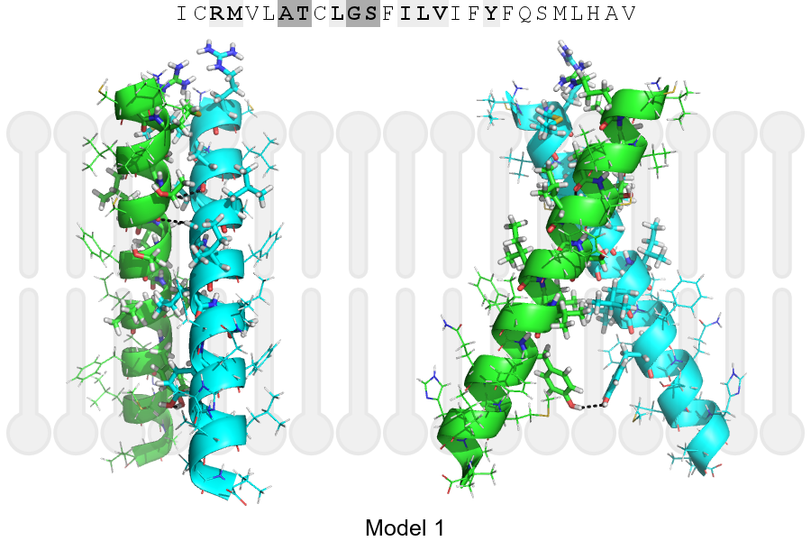
Click on the image to enlarge
Download model 1 structure: PDB, PyMOL session
List of hydrogen bonds:
| Donor | Acceptor | Distance |
|---|---|---|
| B, 21, THR, HA | A, 20, ALA, O | 2.77Å |
| B, 24, GLY, HA1 | A, 21, THR, O | 2.82Å |
| A, 21, THR, HA | B, 20, ALA, O | 2.77Å |
| A, 24, GLY, HA1 | B, 21, THR, O | 2.82Å |
| A, 32, TYR, HH | B, 32, TYR, OH | 2.35Å |