

UniProt data
UniProt name: ILRL2_HUMAN (Q9HB29)
Gene name: IL1RL2; Synonyms=IL1RRP2
Organism: Homo sapiens (Human).
Function: Receptor for interleukin-36 (IL36A, IL36B and IL36G). After binding to interleukin-36 associates with the coreceptor IL1RAP to form the interleukin-36 receptor complex which mediates interleukin-36-dependent activation of NF-kappa-B, MAPK and other pathways (By similarity). The IL-36 signaling system is thought to be present in epithelial barriers and to take part in local inflammatory response; it is similar to the IL-1 system. Seems to be involved in skin inflammatory response by induction of the IL- 23/IL-17/IL-22 pathway. {ECO:0000250|UniProtKB:Q9ERS7, ECO:0000269|PubMed:11466363}.
Subcellular location: Membrane; Single-pass type I membrane protein {ECO:0000269|PubMed:24829417}.
Transmembrane domains:
- 336-356, 21 AA, Helical. {ECO:0000255}.
Sequence: 575 AA
CATM prediction
The sequence of ILRL2_HUMAN returned 1 potential models
The positions at the dimer's interface are highlighted in yellow. The position at the crossing point is highlighted in orange.
| # | Sequence and interface | d | θ | Z' | ω' | Score |
|---|---|---|---|---|---|---|
| 340 350 + | + | + |
(Å) | (°) | (Å) | (°) | ||
| Model 1 | DFRAYLIGGLIALVAVAVSVVYIYNIF | 6.5 | -53.0° | 1.9 | 45.4 | -31.9 |
Click on the model number to visualize its details below.
MODEL 1 DETAILS
Precited structure:
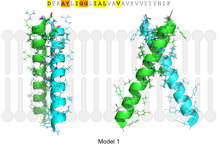
Click on the image to enlarge
Download model 1 structure: PDB, PyMOL session
List of hydrogen bonds:
| Donor | Acceptor | Distance |
|---|---|---|
| B, 337, TYR, HA | A, 336, ALA, O | 2.98Å |
| B, 340, GLY, HA1 | A, 337, TYR, O | 2.42Å |
| B, 341, GLY, HA2 | A, 340, GLY, O | 2.84Å |
| A, 337, TYR, HA | B, 336, ALA, O | 2.98Å |
| A, 340, GLY, HA1 | B, 337, TYR, O | 2.42Å |
| A, 341, GLY, HA2 | B, 340, GLY, O | 2.84Å |