UniProt data
UniProt name: CADM1_HUMAN (Q9BY67)
Gene name: CADM1 {ECO:0000312|HGNC:HGNC:5951}
Organism: Homo sapiens (Human).
Function: Mediates homophilic cell-cell adhesion in a Ca(2+)- independent manner. Also mediates heterophilic cell-cell adhesion with CADM3 and NECTIN3 in a Ca(2+)-independent manner. Acts as a tumor suppressor in non-small-cell lung cancer (NSCLC) cells. Interaction with CRTAM promotes natural killer (NK) cell cytotoxicity and interferon-gamma (IFN-gamma) secretion by CD8+ cells in vitro as well as NK cell-mediated rejection of tumors expressing CADM3 in vivo. May contribute to the less invasive phenotypes of lepidic growth tumor cells. In mast cells, may mediate attachment to and promote communication with nerves. CADM1, together with MITF, is essential for development and survival of mast cells in vivo. Acts as a synaptic cell adhesion molecule and plays a role in the formation of dendritic spines and in synapse assembly (By similarity). May be involved in neuronal migration, axon growth, pathfinding, and fasciculation on the axons of differentiating neurons. May play diverse roles in the spermatogenesis including in the adhesion of spermatocytes and spermatids to Sertoli cells and for their normal differentiation into mature spermatozoa. {ECO:0000250, ECO:0000269|PubMed:11279526, ECO:0000269|PubMed:12050160, ECO:0000269|PubMed:12234973, ECO:0000269|PubMed:12920246, ECO:0000269|PubMed:15811952, ECO:0000269|PubMed:15905536, ECO:0000269|PubMed:22438059}.
Subcellular location: Cell membrane {ECO:0000269|PubMed:11279526, ECO:0000269|PubMed:22438059}; Single-pass type I membrane protein {ECO:0000269|PubMed:11279526}. Cell junction, synapse {ECO:0000250|UniProtKB:Q8R5M8}. Note=Localized to the basolateral plasma membrane of epithelial cells in gall bladder. {ECO:0000250|UniProtKB:Q8R5M8}.
Transmembrane domains:
- 375-395, 21 AA, Helical. {ECO:0000255}.
Sequence: 442 AA
CATM prediction
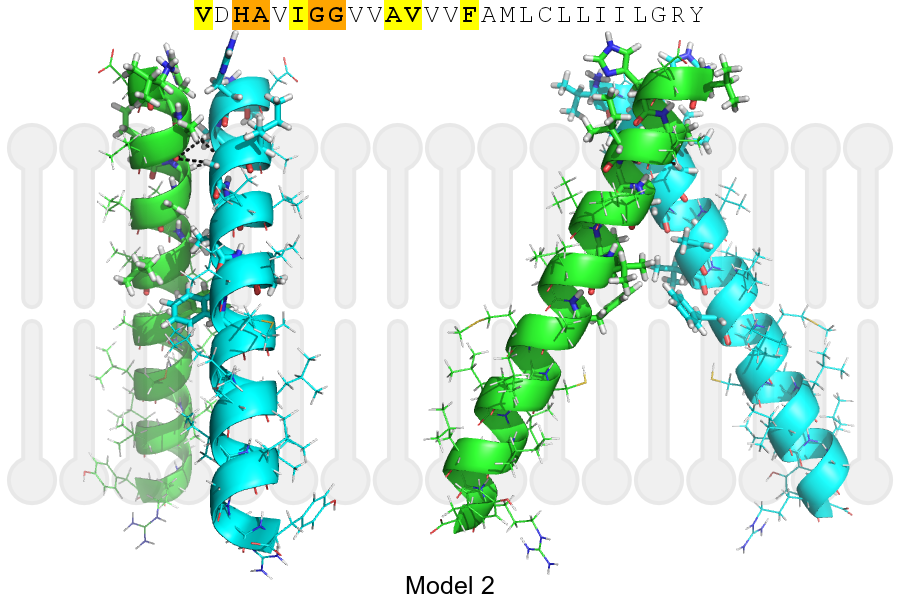
The sequence of CADM1_HUMAN returned 2 potential models
The positions at the dimer's interface are highlighted in yellow. The position at the crossing point is highlighted in orange.
| # | Sequence and interface | d | θ | Z' | ω' | Score |
|---|---|---|---|---|---|---|
| 380 390 + | + | + |
(Å) | (°) | (Å) | (°) | ||
| Model 1 | VDHAVIGGVVAVVVFAMLCLLIILGRY | 6.9 | -43.0° | 4.5 | 35.0 | -29.3 |
| Model 2 | VDHAVIGGVVAVVVFAMLCLLIILGRY | 6.5 | -55.0° | 4.2 | 38.0 | -9.3 |
Click on the model number to visualize its details below.
MODEL 1 DETAILS
Precited structure:
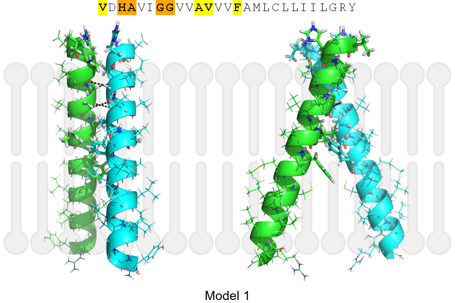
Click on the image to enlarge
Download model 1 structure: PDB, PyMOL session
List of hydrogen bonds:
| Donor | Acceptor | Distance |
|---|---|---|
| B, 378, GLY, HA1 | A, 375, ALA, O | 2.76Å |
| B, 379, GLY, HA2 | A, 378, GLY, O | 3.42Å |
| A, 378, GLY, HA1 | B, 375, ALA, O | 2.76Å |
| A, 379, GLY, HA2 | B, 378, GLY, O | 3.42Å |