UniProt data
UniProt name: PINK1_HUMAN (Q9BXM7)
Gene name: PINK1
Organism: Homo sapiens (Human).
Function: Protects against mitochondrial dysfunction during cellular stress by phosphorylating mitochondrial proteins. Involved in the clearance of damaged mitochondria via selective autophagy (mitophagy) by mediating activation and translocation of PARK2. Targets PARK2 to dysfunctional depolarized mitochondria through the phosphorylation of MFN2. Activates PARK2 in 2 steps: (1) by mediating phosphorylation at 'Ser-65' of PARK2 and (2) mediating phosphorylation of ubiquitin, converting PARK2 to its fully-active form (PubMed:24660806, PubMed:24751536, PubMed:24784582, PubMed:25527291). {ECO:0000269|PubMed:14607334, ECO:0000269|PubMed:15087508, ECO:0000269|PubMed:19229105, ECO:0000269|PubMed:19966284, ECO:0000269|PubMed:20404107, ECO:0000269|PubMed:20798600, ECO:0000269|PubMed:23620051, ECO:0000269|PubMed:23754282, ECO:0000269|PubMed:23933751, ECO:0000269|PubMed:24660806, ECO:0000269|PubMed:24751536, ECO:0000269|PubMed:24784582, ECO:0000269|PubMed:24896179, ECO:0000269|PubMed:25527291}.
Subcellular location: Mitochondrion outer membrane; Single-pass membrane protein {ECO:0000269|PubMed:15087508, ECO:0000269|PubMed:18687899, ECO:0000269|PubMed:19229105, ECO:0000269|PubMed:20798600, ECO:0000269|PubMed:23933751}. Cytoplasm, cytosol {ECO:0000269|PubMed:19229105, ECO:0000269|PubMed:20798600}. Note=Localizes mostly in mitochondrion and the 2 proteolytic processed fragments of 55 kDa and 48 kDa localize mainly in cytosol. {ECO:0000269|PubMed:19229105}.
Transmembrane domains:
- 94-110, 17 AA, Helical. {ECO:0000255}.
Sequence: 581 AA
CATM prediction
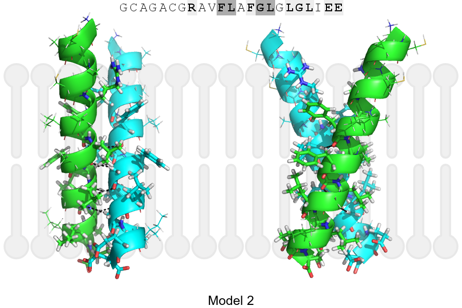
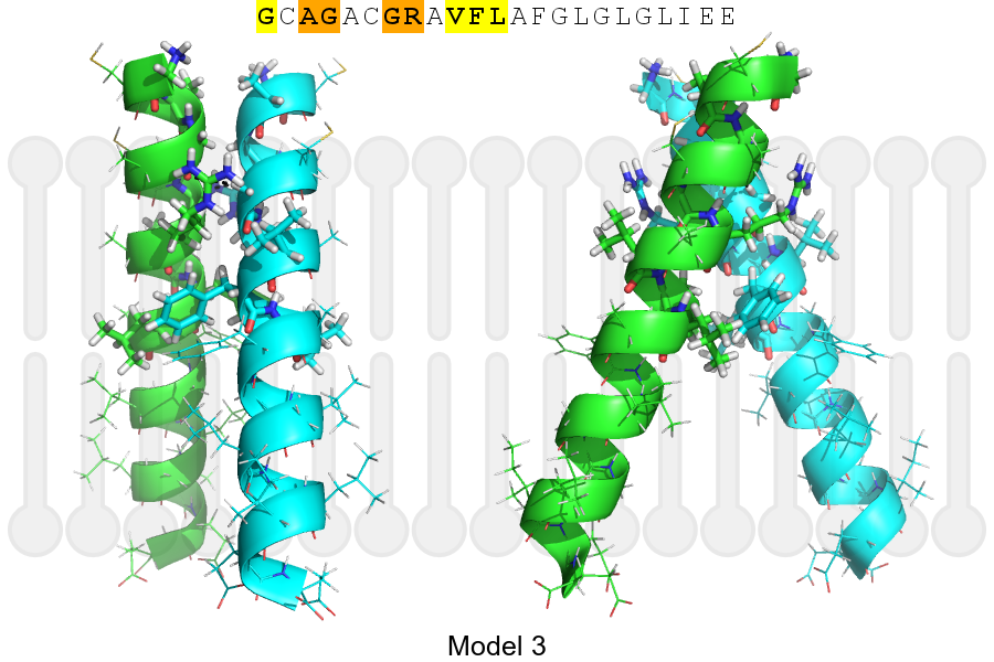
The sequence of PINK1_HUMAN returned 3 potential models
The positions at the dimer's interface are highlighted in yellow. The position at the crossing point is highlighted in orange.
| # | Sequence and interface | d | θ | Z' | ω' | Score |
|---|---|---|---|---|---|---|
| 100 110 + | + | |
(Å) | (°) | (Å) | (°) | ||
| Model 1 | GCAGACGRAVFLAFGLGLGLIEE | 7.2 | -28.0° | 2.4 | 49.9 | -32.5 |
| Model 2 | GCAGACGRAVFLAFGLGLGLIEE | 6.6 | -45.0° | 0.2 | 44.9 | -16.4 |
| Model 3 | GCAGACGRAVFLAFGLGLGLIEE | 7.0 | -46.0° | 3.6 | 37.0 | -15.3 |
| Prolines* | GCAGPCGRAVFLAFGLGLGLIEE |
* Note: this sequence contains Pro residues. CATM substituted them with Ala during the modeling. The positions are marked.
Click on the model number to visualize its details below.
MODEL 1 DETAILS
Precited structure:
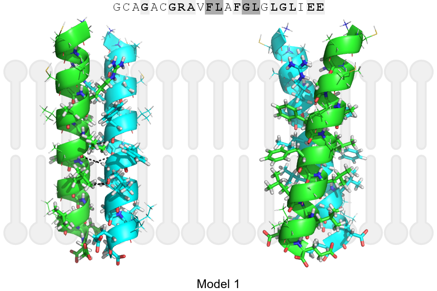
Click on the image to enlarge
Download model 1 structure: PDB, PyMOL session
List of hydrogen bonds:
| Donor | Acceptor | Distance |
|---|---|---|
| B, 102, LEU, HA | A, 101, PHE, O | 3.08Å |
| B, 105, GLY, HA1 | A, 102, LEU, O | 3.33Å |
| B, 106, LEU, HA | A, 105, GLY, O | 3.43Å |
| A, 102, LEU, HA | B, 101, PHE, O | 3.08Å |
| A, 105, GLY, HA1 | B, 102, LEU, O | 3.33Å |
| A, 106, LEU, HA | B, 105, GLY, O | 3.43Å |