UniProt data
UniProt name: JAM3_HUMAN (Q9BX67)
Gene name: JAM3; ORFNames=UNQ859/PRO1868
Organism: Homo sapiens (Human).
Function: Participates in cell-cell adhesion. It is a counter- receptor for ITGAM, mediating leukocyte-platelet interactions and is involved in the regulation of transepithelial migration of polymorphonuclear neutrophils (PMN). The soluble form is a mediator of angiogenesis. {ECO:0000269|PubMed:12208882, ECO:0000269|PubMed:15194813, ECO:0000269|PubMed:20592283}.
Subcellular location: Cell membrane {ECO:0000305}; Single-pass type I membrane protein {ECO:0000305}. Cell junction, desmosome {ECO:0000269|PubMed:15194813}. Secreted, extracellular space {ECO:0000269|PubMed:20592283}. Note=In epithelial cells, it is expressed at desmosomes but not at tight junctions (PubMed:15194813). Localizes at the cell surface of endothelial cells; treatment of endothelial cells with vascular endothelial growth factor stimulates recruitment of JAM3 to cell-cell contacts (PubMed:15994945).
Transmembrane domains:
- 242-262, 21 AA, Helical. {ECO:0000255}.
Sequence: 310 AA
CATM prediction
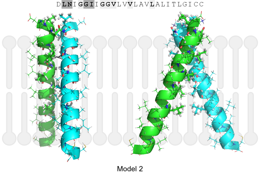
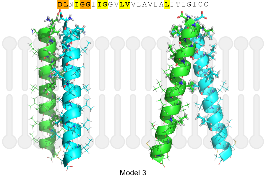
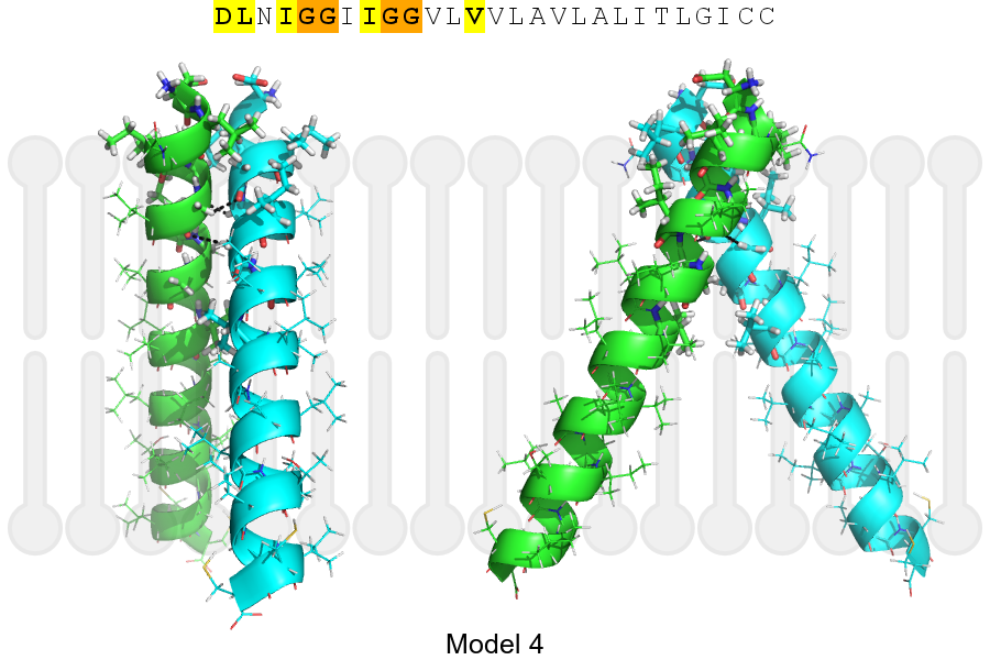
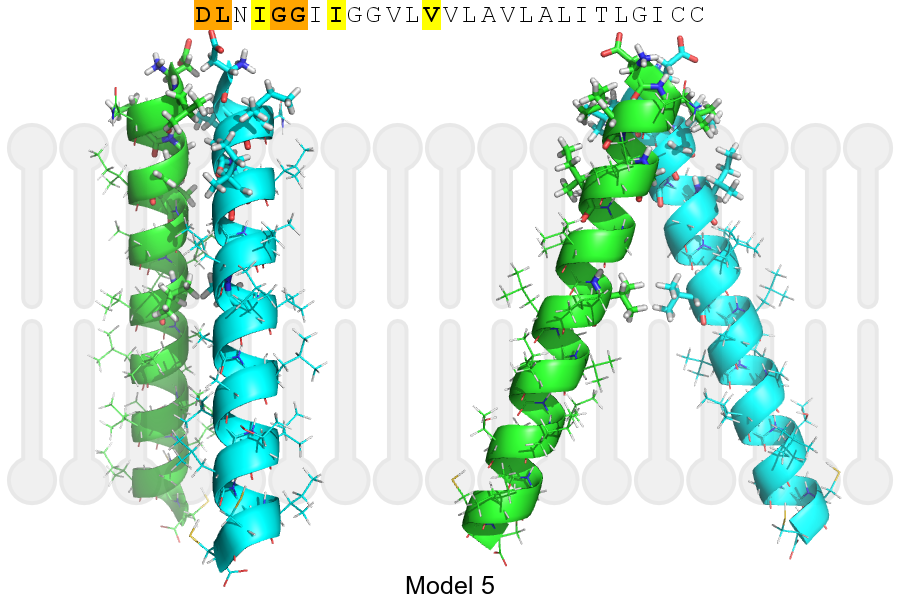
The sequence of JAM3_HUMAN returned 5 potential models
The positions at the dimer's interface are highlighted in yellow. The position at the crossing point is highlighted in orange.
| # | Sequence and interface | d | θ | Z' | ω' | Score |
|---|---|---|---|---|---|---|
| 240 250 260 | + | + | + |
(Å) | (°) | (Å) | (°) | ||
| Model 1 | DLNIGGIIGGVLVVLAVLALITLGICC | 6.5 | -45.1° | 4.0 | 39.0 | -19.5 |
| Model 2 | DLNIGGIIGGVLVVLAVLALITLGICC | 6.6 | -48.0° | 0.8 | 54.0 | -15.1 |
| Model 3 | DLNIGGIIGGVLVVLAVLALITLGICC | 6.8 | -28.0° | 6.0 | 47.0 | -9.0 |
| Model 4 | DLNIGGIIGGVLVVLAVLALITLGICC | 6.4 | -55.0° | 4.9 | 53.3 | -8.7 |
| Model 5 | DLNIGGIIGGVLVVLAVLALITLGICC | 7.0 | -40.0° | 5.1 | 44.0 | -6.5 |
Click on the model number to visualize its details below.
MODEL 1 DETAILS
Precited structure:
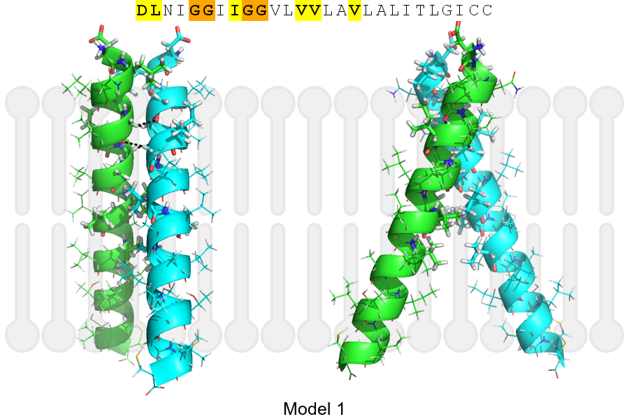
Click on the image to enlarge
Download model 1 structure: PDB, PyMOL session
List of hydrogen bonds:
| Donor | Acceptor | Distance |
|---|---|---|
| B, 244, GLY, HA2 | A, 243, GLY, O | 2.51Å |
| B, 247, GLY, HA1 | A, 244, GLY, O | 2.70Å |
| A, 244, GLY, HA2 | B, 243, GLY, O | 2.51Å |
| A, 247, GLY, HA1 | B, 244, GLY, O | 2.70Å |