

UniProt data
UniProt name: TMED9_HUMAN (Q9BVK6)
Gene name: TMED9; Synonyms=GP25L2
Organism: Homo sapiens (Human).
Function: Appears to be involved in vesicular protein trafficking, mainly in the early secretory pathway. In COPI vesicle-mediated retrograde transport involved in the coatomer recruitment to membranes of the early secretory pathway. Increases coatomer- dependent activity of ARFGAP2. Thought to play a crucial role in the specific retention of p24 complexes in cis-Golgi membranes; specifically contributes to the coupled localization of TMED2 and TMED10 in the cis-Golgi network. May be involved in organization of intracellular membranes, such as of the ER-Golgi intermediate compartment and the Golgi apparatus. Involved in ER localization of PTPN2 isoform PTPB. {ECO:0000269|PubMed:10852829, ECO:0000269|PubMed:14600267, ECO:0000269|PubMed:16595549, ECO:0000269|PubMed:18287528, ECO:0000269|PubMed:19296914}.
Subcellular location: Endoplasmic reticulum membrane; Single-pass type I membrane protein. Golgi apparatus, cis-Golgi network membrane; Single-pass type I membrane protein. Endoplasmic reticulum-Golgi intermediate compartment membrane; Single-pass type I membrane protein. Golgi apparatus, trans-Golgi network membrane {ECO:0000250}; Single-pass type I membrane protein {ECO:0000250}. Note=Cycles between compartments of the early secretatory pathway.
Transmembrane domains:
- 203-222, 20 AA, Helical. {ECO:0000255}.
Sequence: 235 AA
CATM prediction
The sequence of TMED9_HUMAN returned 1 potential models
The positions at the dimer's interface are highlighted in yellow. The position at the crossing point is highlighted in orange.
| # | Sequence and interface | d | θ | Z' | ω' | Score |
|---|---|---|---|---|---|---|
| 200 210 220 | + | + | + |
(Å) | (°) | (Å) | (°) | ||
| Model 1 | NQRVLWWSILQTLILVAIGVWQMRHL | 7.3 | -51.0° | 2.4 | 44.9 | -7.1 |
Click on the model number to visualize its details below.
MODEL 1 DETAILS
Precited structure:
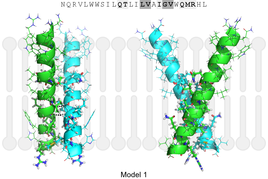
Click on the image to enlarge
Download model 1 structure: PDB, PyMOL session
List of hydrogen bonds:
| Donor | Acceptor | Distance |
|---|---|---|
| B, 215, VAL, HA | A, 214, LEU, O | 3.42Å |
| B, 218, GLY, HA1 | A, 215, VAL, O | 3.22Å |
| A, 215, VAL, HA | B, 214, LEU, O | 3.42Å |
| A, 218, GLY, HA1 | B, 215, VAL, O | 3.22Å |