

UniProt data
UniProt name: MIC26_HUMAN (Q9BUR5)
Gene name: APOO
Organism: Homo sapiens (Human).
Function: Component of the MICOS complex, a large protein complex of the mitochondrial inner membrane that plays crucial roles in the maintenance of crista junctions, inner membrane architecture, and formation of contact sites to the outer membrane. Plays a crucial role in crista junction formation and mitochondrial function (PubMed:25764979). Can promote cardiac lipotoxicity by enhancing mitochondrial respiration and fatty acid metabolism in cardiac myoblasts (PubMed:24743151). Promotes cholesterol efflux from macrophage cells. Detected in HDL, LDL and VLDL. Secreted by a microsomal triglyceride transfer protein (MTTP)-dependent mechanism, probably as a VLDL-associated protein that is subsequently transferred to HDL (PubMed:16956892). {ECO:0000269|PubMed:16956892, ECO:0000269|PubMed:24743151, ECO:0000269|PubMed:25764979}.
Subcellular location: Mitochondrion inner membrane {ECO:0000269|PubMed:25764979}; Single-pass membrane protein {ECO:0000255}. Secreted {ECO:0000269|PubMed:16956892, ECO:0000269|PubMed:25764979}. Mitochondrion {ECO:0000269|PubMed:25781180}. Golgi apparatus membrane {ECO:0000269|PubMed:25764979}. Endoplasmic reticulum membrane {ECO:0000269|PubMed:25764979}. Note=Exists in three distinct forms: a glycosylated and secreted form, an ER/Golgi-resident form and a non-glycosylated mitochondrial form. {ECO:0000269|PubMed:25764979}.
Transmembrane domains:
- 108-128, 21 AA, Helical. {ECO:0000255}.
Sequence: 198 AA
CATM prediction
The sequence of MIC26_HUMAN returned 1 potential models
The positions at the dimer's interface are highlighted in yellow. The position at the crossing point is highlighted in orange.
| # | Sequence and interface | d | θ | Z' | ω' | Score |
|---|---|---|---|---|---|---|
| 110 120 13 + | + | + | |
(Å) | (°) | (Å) | (°) | ||
| Model 1 | NAAAGFFARLGVIGFAGLIGLLLARGS | 6.8 | -45.8° | 2.7 | 47.0 | -21.7 |
| Prolines* | NAPPGFFPRLGVIGFAGLIGLLLARGS |
* Note: this sequence contains Pro residues. CATM substituted them with Ala during the modeling. The positions are marked.
Click on the model number to visualize its details below.
MODEL 1 DETAILS
Precited structure:
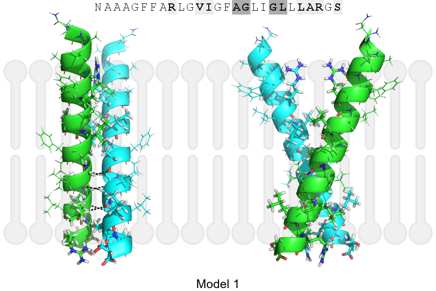
Click on the image to enlarge
Download model 1 structure: PDB, PyMOL session
List of hydrogen bonds:
| Donor | Acceptor | Distance |
|---|---|---|
| B, 121, GLY, HA2 | A, 120, ALA, O | 2.83Å |
| B, 124, GLY, HA1 | A, 121, GLY, O | 2.78Å |
| B, 125, LEU, HA | A, 124, GLY, O | 3.36Å |
| A, 121, GLY, HA2 | B, 120, ALA, O | 2.83Å |
| A, 124, GLY, HA1 | B, 121, GLY, O | 2.78Å |
| A, 125, LEU, HA | B, 124, GLY, O | 3.36Å |