

UniProt data
UniProt name: MEG10_HUMAN (Q96KG7)
Gene name: MEGF10; Synonyms=KIAA1780
Organism: Homo sapiens (Human).
Function: Membrane receptor involved in phagocytosis by macrophages of apoptotic cells. Cooperates with ABCA1 within the process of engulfment. Promotes the formation of large intracellular vacuoles and may be responsible for the uptake of amyloid-beta peptides. May also function in the mosaic spacing of specific neuron subtypes in the retina through homotypic retinal neuron repulsion. Mosaics provide a mechanism to distribute each cell type evenly across the retina, ensuring that all parts of the visual field have access to a full set of processing elements. May play role in cell adhesion and motility. Is also an essential factor in the regulation of myogenesis. Controls the balance between skeletal muscle satellite cells proliferation and differentiation probably through regulation of the notch signaling pathway. {ECO:0000269|PubMed:17498693, ECO:0000269|PubMed:17643423, ECO:0000269|PubMed:20828568, ECO:0000269|PubMed:22101682}.
Subcellular location: Cell membrane; Single-pass type I membrane protein. Basolateral cell membrane; Single-pass type I membrane protein. Cell projection, phagocytic cup. Note=Enriched at the sites of contact with apoptotic thymocyte cells (By similarity). Forms an irregular, mosaic-like adhesion pattern in region of the cell surface that becomes firmely fixed to the substrate. Expressed at the cell surface in clusters around cell corpses during engulfment. During the engulfment of apoptotic thymocytes, recruited at the bottom of the forming phagocytic cup. Colocalizes with ABCA1 in absence of any phagocytic challenge. Does not localize within lamellipodia. Does not localize with MEGF11. {ECO:0000250}.
Transmembrane domains:
- 858-878, 21 AA, Helical. {ECO:0000255}.
Sequence: 1140 AA
CATM prediction
The sequence of MEG10_HUMAN returned 1 potential models
The positions at the dimer's interface are highlighted in yellow. The position at the crossing point is highlighted in orange.
| # | Sequence and interface | d | θ | Z' | ω' | Score |
|---|---|---|---|---|---|---|
| 860 870 88 + | + | + | |
(Å) | (°) | (Å) | (°) | ||
| Model 1 | QIGAIAGIIILVLVVLFLLALFIIYRH | 6.6 | -55.0° | 5.6 | 39.9 | -13.1 |
Click on the model number to visualize its details below.
MODEL 1 DETAILS
Precited structure:
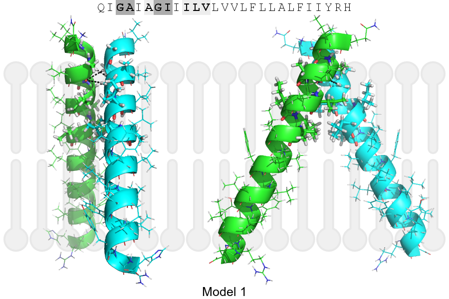
Click on the image to enlarge
Download model 1 structure: PDB, PyMOL session
List of hydrogen bonds:
| Donor | Acceptor | Distance |
|---|---|---|
| B, 858, ALA, HA | A, 858, ALA, O | 3.49Å |
| B, 861, GLY, HA1 | A, 858, ALA, O | 3.16Å |
| A, 858, ALA, HA | B, 858, ALA, O | 3.49Å |
| A, 861, GLY, HA1 | B, 858, ALA, O | 3.16Å |