UniProt data
UniProt name: PGAM5_HUMAN (Q96HS1)
Gene name: PGAM5
Organism: Homo sapiens (Human).
Function: Displays phosphatase activity for serine/threonine residues, and, dephosphorylates and activates MAP3K5 kinase. Has apparently no phosphoglycerate mutase activity. May be regulator of mitochondrial dynamics. Substrate for a KEAP1-dependent ubiquitin ligase complex. Contributes to the repression of NFE2L2- dependent gene expression. Acts as a central mediator for programmed necrosis induced by TNF, by reactive oxygen species and by calcium ionophore. {ECO:0000269|PubMed:18387606, ECO:0000269|PubMed:19590015, ECO:0000269|PubMed:22265414}.
Subcellular location: Mitochondrion outer membrane {ECO:0000269|PubMed:18387606}; Single-pass membrane protein {ECO:0000269|PubMed:18387606}. Note=Isoform 2 overexpression results in the formation of disconnected punctuate mitochondria distributed throughout the cytoplasm. Isoform 1 overexpression results in the clustering of mitochondria around the nucleus.
Transmembrane domains:
- 7-29, 23 AA, Helical. {ECO:0000255}.
Sequence: 289 AA
CATM prediction
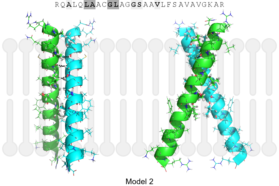
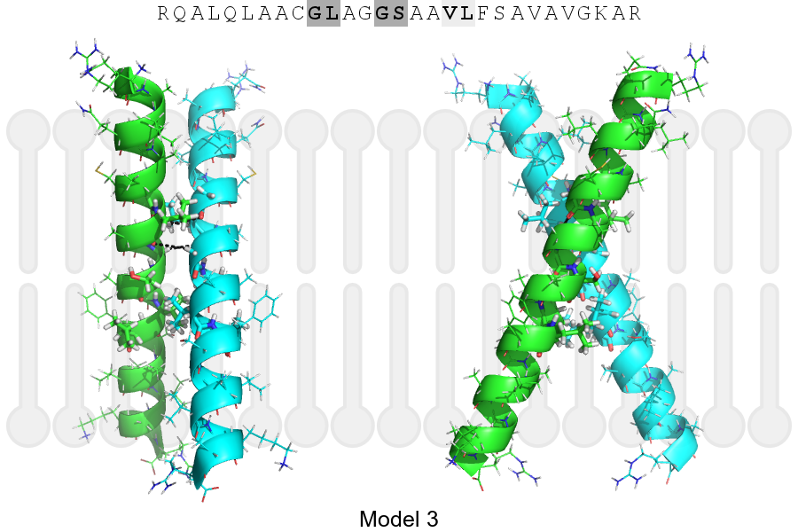
The sequence of PGAM5_HUMAN returned 3 potential models
The positions at the dimer's interface are highlighted in yellow. The position at the crossing point is highlighted in orange.
| # | Sequence and interface | d | θ | Z' | ω' | Score |
|---|---|---|---|---|---|---|
| 10 20 30 + | + | + | |
(Å) | (°) | (Å) | (°) | ||
| Model 1 | RQALQLAACGLAGGSAAVLFSAVAVGKAR | 6.8 | -42.7° | 3.0 | 34.9 | -26.0 |
| Model 2 | RQALQLAACGLAGGSAAVLFSAVAVGKAR | 6.8 | -47.1° | 1.3 | 45.0 | -17.8 |
| Model 3 | RQALQLAACGLAGGSAAVLFSAVAVGKAR | 7.4 | -49.9° | 2.9 | 46.8 | -3.8 |
| Prolines* | RQALQLAACGLAGGSAAVLFSAVAVGKPR |
* Note: this sequence contains Pro residues. CATM substituted them with Ala during the modeling. The positions are marked.
Click on the model number to visualize its details below.
MODEL 1 DETAILS
Precited structure:
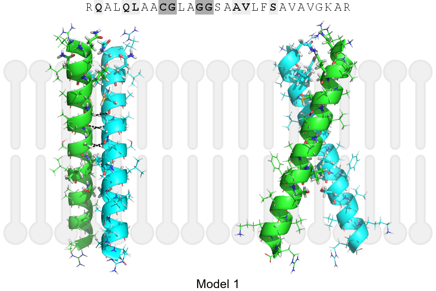
Click on the image to enlarge
Download model 1 structure: PDB, PyMOL session
List of hydrogen bonds:
| Donor | Acceptor | Distance |
|---|---|---|
| B, 13, GLY, HA2 | A, 12, CYS, O | 2.95Å |
| B, 16, GLY, HA1 | A, 13, GLY, O | 2.76Å |
| B, 17, GLY, HA2 | A, 16, GLY, O | 3.10Å |
| A, 13, GLY, HA2 | B, 12, CYS, O | 2.95Å |
| A, 16, GLY, HA1 | B, 13, GLY, O | 2.76Å |
| A, 17, GLY, HA2 | B, 16, GLY, O | 3.10Å |