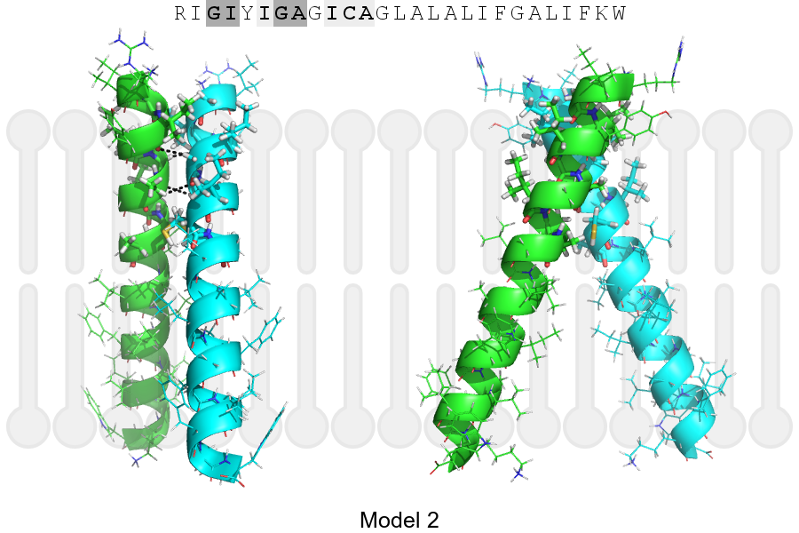
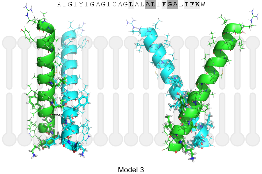
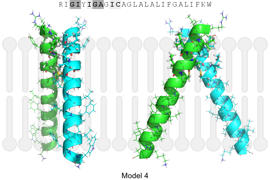
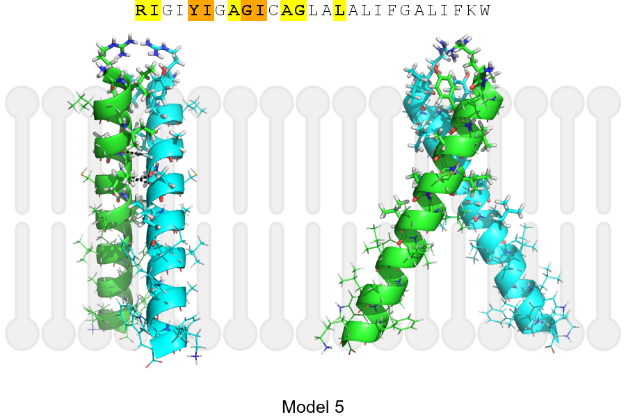
UniProt data
UniProt name: HAVR2_HUMAN (Q8TDQ0)
Gene name: HAVCR2; Synonyms=TIM3, TIMD3
Organism: Homo sapiens (Human).
Function: Cell surface receptor implicated in modulating innate and adaptive immune responses. Generally accepted to have an inhibiting function. Reports on stimulating functions suggest that the activity may be influenced by the cellular context and/or the respective ligand (PubMed:24825777). Regulates macrophage activation (PubMed:11823861). Inhibits T-helper type 1 lymphocyte (Th1)-mediated auto- and alloimmune responses and promotes immunological tolerance (PubMed:14556005). In CD8+ cells attenuates TCR-induced signaling, specifically by blocking NF- kappaB and NFAT promoter activities resulting in the loss of IL-2 secretion. The function may implicate its association with LCK proposed to impair phosphorylation of TCR subunits, and/or LGALS9- dependent recruitment of PTPRC to the immunological synapse (PubMed:24337741, PubMed:26492563). In contrast, shown to activate TCR-induced signaling in T-cells probably implicating ZAP70, LCP2, LCK and FYN (By similarity). Expressed on Treg cells can inhibit Th17 cell responses (PubMed:24838857). Receptor for LGALS9 (PubMed:16286920, PubMed:24337741). Binding to LGALS9 is believed to result in suppression of T-cell responses; the resulting apoptosis of antigen-specific cells may implicate HAVCR2 phosphorylation and disruption of its association with BAG6. Binding to LGALS9 is proposed to be involved in innate immune response to intracellular pathogens. Expressed on Th1 cells interacts with LGALS9 expressed on Mycobacterium tuberculosis- infected macrophages to stimulate antibactericidal activity including IL-1 beta secretion and to restrict intracellular bacterial growth (By similarity). However, the function as receptor for LGALS9 has been challenged (PubMed:23555261). Also reported to enhance CD8+ T-cell responses to an acute infection such as by Listeria monocytogenes (By similarity). Receptor for phosphatidylserine (PtSer); PtSer-binding is calcium-dependent. May recognize PtSer on apoptotic cells leading to their phagocytosis. Mediates the engulfment of apoptotic cells by dendritic cells. Expressed on T-cells, promotes conjugation but not engulfment of apoptotic cells. Expressed on dendritic cells (DCs) positively regulates innate immune response and in synergy with Toll-like receptors promotes secretion of TNF-alpha. In tumor-imfiltrating DCs suppresses nucleic acid-mediated innate immune repsonse by interaction with HMGB1 and interfering with nucleic acid-sensing and trafficking of nucleid acids to endosomes (By similarity). Expressed on natural killer (NK) cells acts as a coreceptor to enhance IFN-gamma production in response to LGALS9 (PubMed:22323453). In contrast, shown to suppress NK cell-mediated cytotoxicity (PubMed:22383801). Negatively regulates NK cell function in LPS-induced endotoxic shock (By similarity). {ECO:0000250|UniProtKB:Q8VIM0, ECO:0000269|PubMed:11823861, ECO:0000269|PubMed:14556005, ECO:0000269|PubMed:16286920, ECO:0000269|PubMed:22323453, ECO:0000269|PubMed:23555261, ECO:0000269|PubMed:24838857, ECO:0000269|PubMed:26492563, ECO:0000305|PubMed:24825777}.
Subcellular location: Membrane {ECO:0000305}; Single-pass type I membrane protein {ECO:0000305}. Cell junction {ECO:0000269|PubMed:24337741}. Note=Localizes to the immunological synapse between CD8+ T-cells and target cells. {ECO:0000269|PubMed:24337741}.
Transmembrane domains:
- 203-223, 21 AA, Helical. {ECO:0000255}.
Sequence: 301 AA
CATM prediction
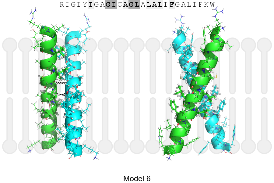
The sequence of HAVR2_HUMAN returned 6 potential models
The positions at the dimer's interface are highlighted in yellow. The position at the crossing point is highlighted in orange.
| # | Sequence and interface | d | θ | Z' | ω' | Score |
|---|---|---|---|---|---|---|
| 200 210 220 | + | + | + |
(Å) | (°) | (Å) | (°) | ||
| Model 1 | RIGIYIGAGICAGLALALIFGALIFKW | 6.4 | -44.9° | 4.3 | 55.0 | -44.8 |
| Model 2 | RIGIYIGAGICAGLALALIFGALIFKW | 6.7 | -46.9° | 2.3 | 44.8 | -24.6 |
| Model 3 | RIGIYIGAGICAGLALALIFGALIFKW | 6.8 | -55.0° | 2.4 | 48.0 | -20.9 |
| Model 4 | RIGIYIGAGICAGLALALIFGALIFKW | 6.5 | -51.0° | 4.8 | 57.0 | -20.5 |
| Model 5 | RIGIYIGAGICAGLALALIFGALIFKW | 7.1 | -54.8° | 2.4 | 48.1 | -19.1 |
| Model 6 | RIGIYIGAGICAGLALALIFGALIFKW | 7.4 | -44.0° | 0.9 | 46.2 | -9.6 |
Click on the model number to visualize its details below.
MODEL 1 DETAILS
Precited structure:
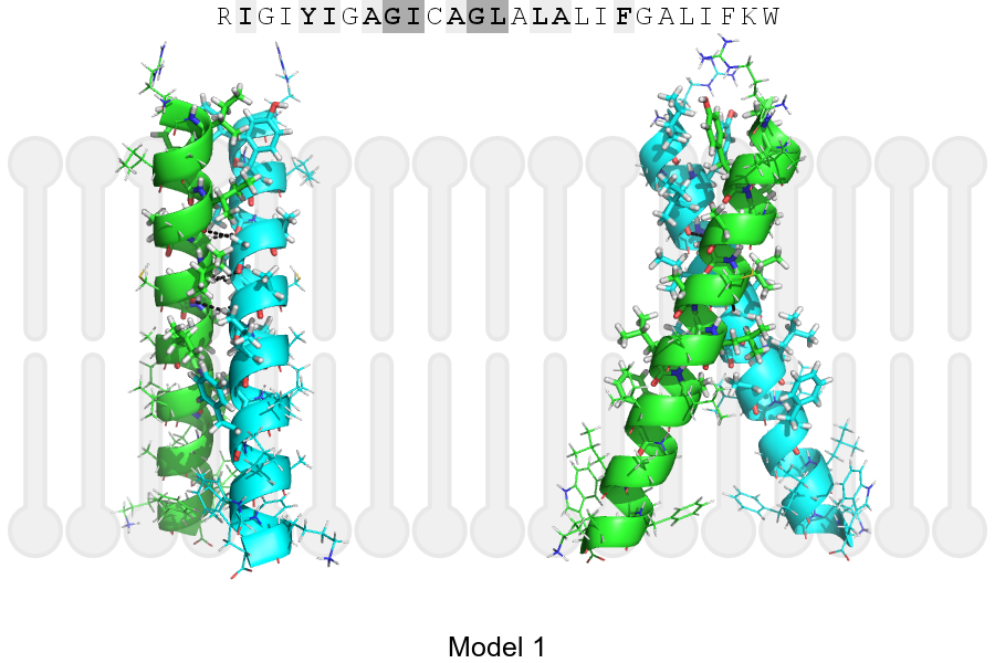
Click on the image to enlarge
Download model 1 structure: PDB, PyMOL session
List of hydrogen bonds:
| Donor | Acceptor | Distance |
|---|---|---|
| B, 208, GLY, HA1 | A, 205, ILE, O | 3.10Å |
| B, 209, ILE, HA | A, 208, GLY, O | 2.35Å |
| B, 212, GLY, HA1 | A, 209, ILE, O | 2.81Å |
| A, 208, GLY, HA1 | B, 205, ILE, O | 3.10Å |
| A, 209, ILE, HA | B, 208, GLY, O | 2.35Å |
| A, 212, GLY, HA1 | B, 209, ILE, O | 2.81Å |