

UniProt data
UniProt name: R144B_HUMAN (Q7Z419)
Gene name: RNF144B; Synonyms=IBRDC2, P53RFP
Organism: Homo sapiens (Human).
Function: E3 ubiquitin-protein ligase which accepts ubiquitin from E2 ubiquitin-conjugating enzymes UBE2L3 and UBE2L6 in the form of a thioester and then directly transfers the ubiquitin to targeted substrates such as LCMT2, thereby promoting their degradation. Induces apoptosis via a p53/TP53-dependent but caspase-independent mechanism. However, its overexpression also produces a decrease of the ubiquitin-dependent stability of BAX, a pro-apoptotic protein, ultimately leading to protection of cell death; But, it is not an anti-apoptotic protein per se. {ECO:0000269|PubMed:12853982, ECO:0000269|PubMed:20300062}.
Subcellular location: Mitochondrion membrane {ECO:0000269|PubMed:20300062}; Single-pass membrane protein {ECO:0000269|PubMed:20300062}. Cytoplasm {ECO:0000269|PubMed:20300062}. Note=Mostly cytosololic, accumulates in submitochondrial domains specifically upon apoptosis induction, in synchrony with BAX activation.
Transmembrane domains:
- 258-278, 21 AA, Helical. {ECO:0000255}.
Sequence: 303 AA
CATM prediction
The sequence of R144B_HUMAN returned 1 potential models
The positions at the dimer's interface are highlighted in yellow. The position at the crossing point is highlighted in orange.
| # | Sequence and interface | d | θ | Z' | ω' | Score |
|---|---|---|---|---|---|---|
| 260 270 28 + | + | + | |
(Å) | (°) | (Å) | (°) | ||
| Model 1 | RTQVVGILVGLGIIALVTSALLLLASA | 7.2 | -47.0° | 1.4 | 45.0 | -12.4 |
| Prolines* | RTQVVGILVGLGIIALVTSPLLLLASP |
* Note: this sequence contains Pro residues. CATM substituted them with Ala during the modeling. The positions are marked.
Click on the model number to visualize its details below.
MODEL 1 DETAILS
Precited structure:
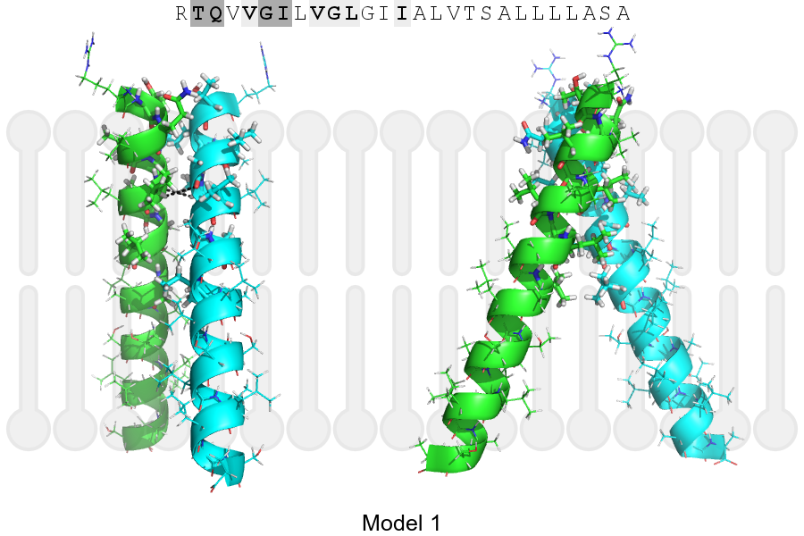
Click on the image to enlarge
Download model 1 structure: PDB, PyMOL session
List of hydrogen bonds:
| Donor | Acceptor | Distance |
|---|---|---|
| B, 261, ILE, HA | A, 260, GLY, O | 3.40Å |
| A, 261, ILE, HA | B, 260, GLY, O | 3.40Å |