UniProt data
UniProt name: CR3L2_HUMAN (Q70SY1)
Gene name: CREB3L2; Synonyms=BBF2H7
Organism: Homo sapiens (Human).
Function: Transcription factor involved in unfolded protein response (UPR). In the absence of endoplasmic reticulum (ER) stress, inserted into ER membranes, with N-terminal DNA-binding and transcription activation domains oriented toward the cytosolic face of the membrane. In response to ER stress, transported to the Golgi, where it is cleaved in a site-specific manner by resident proteases S1P/MBTPS1 and S2P/MBTPS2. The released N-terminal cytosolic domain is translocated to the nucleus to effect transcription of specific target genes. Plays a critical role in chondrogenesis by activating the transcription of SEC23A, which promotes the transport and secretion of cartilage matrix proteins, and possibly that of ER biogenesis-related genes (By similarity). In a neuroblastoma cell line, protects cells from ER stress- induced death (PubMed:17178827). In vitro activates transcription of target genes via direct binding to the CRE site (PubMed:17178827). {ECO:0000250|UniProtKB:Q8BH52, ECO:0000269|PubMed:17178827}.
Subcellular location: Processed cyclic AMP-responsive element- binding protein 3-like protein 2: Nucleus. Note=Upon ER stress, translocated into the nucleus. {ECO:0000250|UniProtKB:Q8BH52}.
Transmembrane domains:
- 380-400, 21 AA, Helical; Signal-anchor for type II membrane protein. {ECO:0000255}.
Sequence: 520 AA
CATM prediction
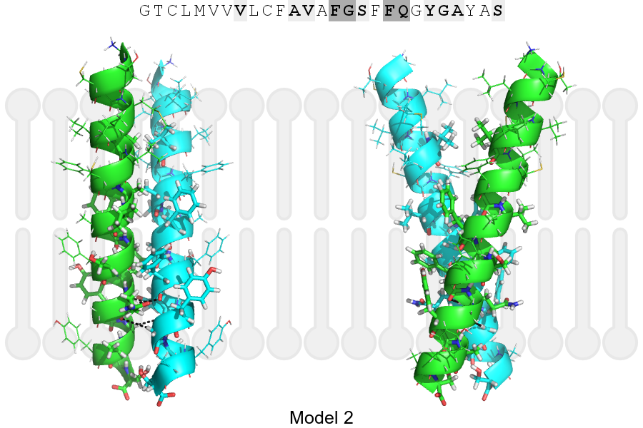
The sequence of CR3L2_HUMAN returned 2 potential models
The positions at the dimer's interface are highlighted in yellow. The position at the crossing point is highlighted in orange.
| # | Sequence and interface | d | θ | Z' | ω' | Score |
|---|---|---|---|---|---|---|
| 380 390 400 | + | + | |
(Å) | (°) | (Å) | (°) | ||
| Model 1 | GTCLMVVVLCFAVAFGSFFQGYGAYAS | 6.9 | -48.8° | 1.8 | 45.1 | -23.5 |
| Model 2 | GTCLMVVVLCFAVAFGSFFQGYGAYAS | 7.0 | -36.9° | 0.0 | 36.8 | -10.1 |
| Prolines* | GTCLMVVVLCFAVAFGSFFQGYGPYPS |
* Note: this sequence contains Pro residues. CATM substituted them with Ala during the modeling. The positions are marked.
Click on the model number to visualize its details below.
MODEL 1 DETAILS
Precited structure:
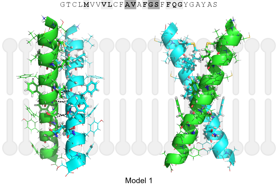
Click on the image to enlarge
Download model 1 structure: PDB, PyMOL session
List of hydrogen bonds:
| Donor | Acceptor | Distance |
|---|---|---|
| B, 389, VAL, HA | A, 388, ALA, O | 3.16Å |
| B, 392, GLY, HA1 | A, 389, VAL, O | 2.84Å |
| B, 393, SER, HA | A, 392, GLY, O | 3.34Å |
| A, 389, VAL, HA | B, 388, ALA, O | 3.16Å |
| A, 392, GLY, HA1 | B, 389, VAL, O | 2.84Å |
| A, 393, SER, HA | B, 392, GLY, O | 3.34Å |