UniProt data
UniProt name: UBP30_HUMAN (Q70CQ3)
Gene name: USP30 {ECO:0000312|HGNC:HGNC:20065}
Organism: Homo sapiens (Human).
Function: Deubiquitinating enzyme tethered to the mitochondrial outer membrane that acts as a key inhibitor of mitophagy by counteracting the action of parkin (PARK2): hydrolyzes ubiquitin attached by parkin on target proteins, such as RHOT1/MIRO1 and TOMM20, thereby blocking parkin's ability to drive mitophagy (PubMed:18287522, PubMed:24896179, PubMed:25527291, PubMed:25621951). Preferentially cleaves 'Lys-6'- and 'Lys-11'- linked polyubiquitin chains, 2 types of linkage that participate to mitophagic signaling (PubMed:25621951). Does not cleave efficiently polyubiquitin phosphorylated at 'Ser-65' (PubMed:25527291). Acts as negative regulator of mitochondrial fusion by mediating deubiquitination of MFN1 and MFN2 (By similarity). {ECO:0000250|UniProtKB:Q3UN04, ECO:0000269|PubMed:18287522, ECO:0000269|PubMed:24896179, ECO:0000269|PubMed:25527291, ECO:0000269|PubMed:25621951}.
Subcellular location: Mitochondrion outer membrane {ECO:0000269|PubMed:18287522, ECO:0000269|PubMed:24896179}.
Transmembrane domains:
- 36-56, 21 AA, Helical. {ECO:0000255}.
Sequence: 517 AA
CATM prediction
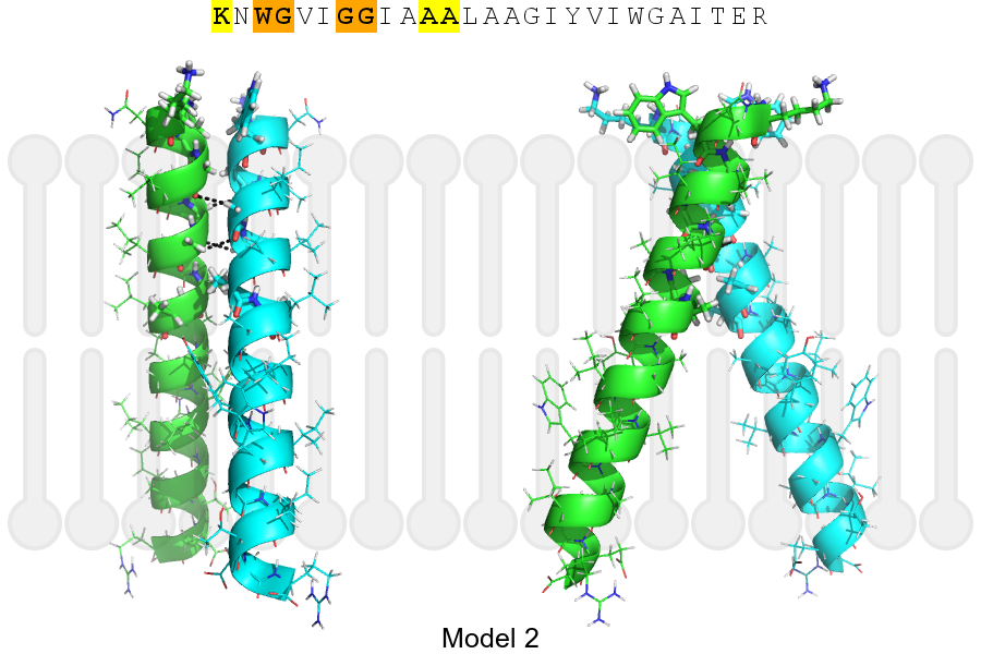
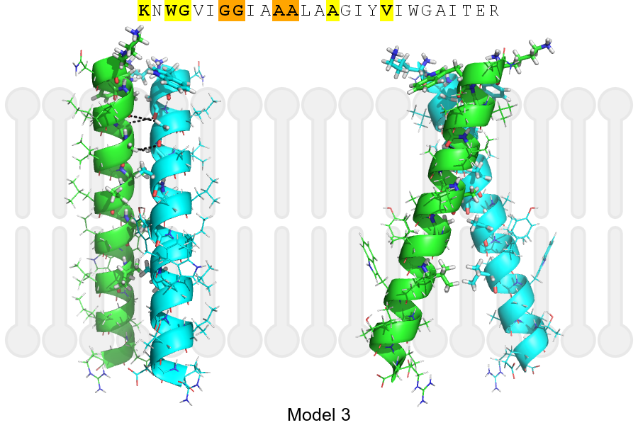
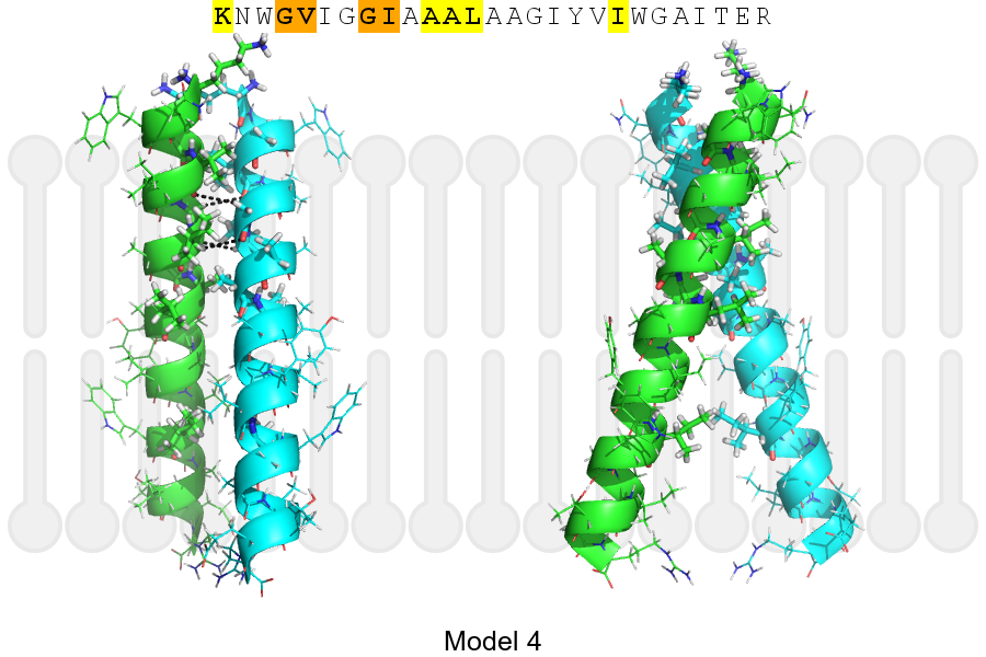
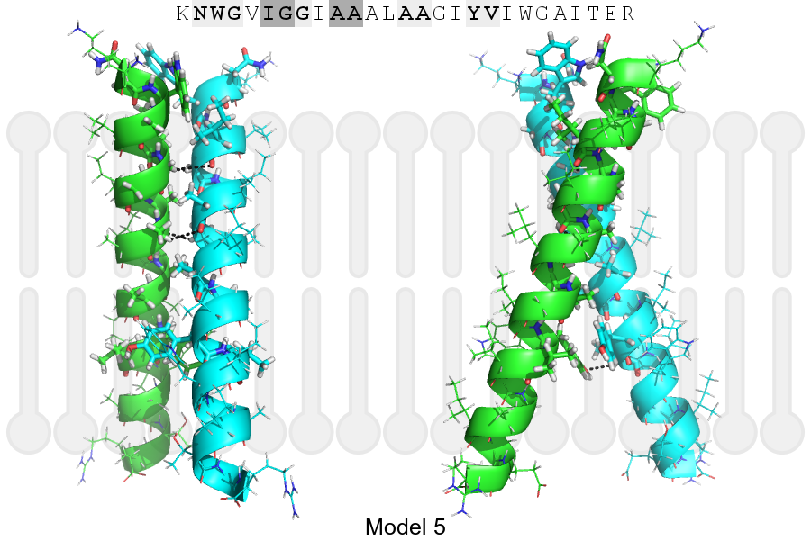
The sequence of UBP30_HUMAN returned 5 potential models
The positions at the dimer's interface are highlighted in yellow. The position at the crossing point is highlighted in orange.
| # | Sequence and interface | d | θ | Z' | ω' | Score |
|---|---|---|---|---|---|---|
| 40 50 + | + | + |
(Å) | (°) | (Å) | (°) | ||
| Model 1 | KNWGVIGGIAAALAAGIYVIWGAITER | 6.4 | -47.1° | 3.3 | 54.3 | -18.1 |
| Model 2 | KNWGVIGGIAAALAAGIYVIWGAITER | 6.6 | -43.0° | 2.4 | 43.9 | -16.3 |
| Model 3 | KNWGVIGGIAAALAAGIYVIWGAITER | 7.0 | -38.0° | 5.7 | 33.7 | -12.2 |
| Model 4 | KNWGVIGGIAAALAAGIYVIWGAITER | 7.4 | -39.2° | 1.1 | 42.9 | -10.3 |
| Model 5 | KNWGVIGGIAAALAAGIYVIWGAITER | 7.0 | -41.9° | 1.9 | 33.1 | -6.5 |
| Prolines* | KNWGVIGGIAAALAAGIYVIWGPITER |
* Note: this sequence contains Pro residues. CATM substituted them with Ala during the modeling. The positions are marked.
Click on the model number to visualize its details below.
MODEL 1 DETAILS
Precited structure:
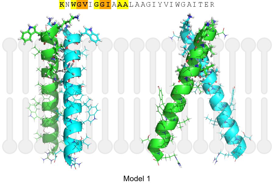
Click on the image to enlarge
Download model 1 structure: PDB, PyMOL session
List of hydrogen bonds:
| Donor | Acceptor | Distance |
|---|---|---|
| B, 37, VAL, HA | A, 36, GLY, O | 2.44Å |
| B, 40, GLY, HA1 | A, 37, VAL, O | 2.58Å |
| B, 41, ILE, HA | A, 40, GLY, O | 3.43Å |
| A, 37, VAL, HA | B, 36, GLY, O | 2.44Å |
| A, 40, GLY, HA1 | B, 37, VAL, O | 2.58Å |
| A, 41, ILE, HA | B, 40, GLY, O | 3.43Å |