UniProt data
UniProt name: 2B1A_HUMAN (Q30167)
Gene name: HLA-DRB1
Organism: Homo sapiens (Human).
Function: Binds peptides derived from antigens that access the endocytic route of antigen presenting cells (APC) and presents them on the cell surface for recognition by the CD4 T-cells. The peptide binding cleft accommodates peptides of 10-30 residues. The peptides presented by MHC class II molecules are generated mostly by degradation of proteins that access the endocytic route; where they are processed by lysosomal proteases and other hydrolases. Exogenous antigens that have been endocytosed by the APC are thus readily available for presentation via MHC II molecules; and for this reason this antigen presentation pathway is usually referred to as exogenous. As membrane proteins on their way to degradation in lysosomes as part of their normal turn-over are also contained in the endosomal/lysosomal compartments; exogenous antigens must compete with those derived from endogenous components. Autophagy is also a source of endogenous peptides; autophagosomes constitutively fuse with MHC class II loading compartments. In addition to APCs; other cells of the gastrointestinal tract; such as epithelial cells; express MHC class II molecules and CD74 and act as APCs; which is an unusual trait of the GI tract. To produce a MHC class II molecule that presents an antigen; three MHC class II molecules (heterodimers of an alpha and a beta chain) associate with a CD74 trimer in the ER to form a heterononamer. Soon after the entry of this complex into the endosomal/lysosomal system where antigen processing occurs; CD74 undergoes a sequential degradation by various proteases; including CTSS and CTSL; leaving a small fragment termed CLIP (class-II-associated invariant chain peptide). The removal of CLIP is facilitated by HLA-DM via direct binding to the alpha-beta-CLIP complex so that CLIP is released. HLA-DM stabilizes MHC class II molecules until primary high affinity antigenic peptides are bound. The MHC II molecule bound to a peptide is then transported to the cell membrane surface. In B-cells; the interaction between HLA-DM and MHC class II molecules is regulated by HLA-DO. Primary dendritic cells (DCs) also to express HLA-DO. Lysosomal microenvironment has been implicated in the regulation of antigen loading into MHC II molecules; increased acidification produces increased proteolysis and efficient peptide loading.
Subcellular location: Cell membrane {ECO:0000269|PubMed:18305173}; Single-pass type I membrane protein {ECO:0000269|PubMed:18305173}. Endoplasmic reticulum membrane {ECO:0000269|PubMed:18305173}; Single-pass type I membrane protein {ECO:0000269|PubMed:18305173}. Golgi apparatus, trans-Golgi network membrane {ECO:0000269|PubMed:18305173}; Single-pass type I membrane protein {ECO:0000269|PubMed:18305173}. Endosome membrane {ECO:0000269|PubMed:18305173}; Single-pass type I membrane protein {ECO:0000269|PubMed:18305173}. Lysosome membrane {ECO:0000269|PubMed:18305173}; Single-pass type I membrane protein {ECO:0000269|PubMed:18305173}. Late endosome membrane {ECO:0000269|PubMed:18305173}; Single-pass type I membrane protein {ECO:0000269|PubMed:18305173}. Note=The MHC class II complex transits through a number of intracellular compartments in the endocytic pathway until it reaches the cell membrane for antigen presentation.
Transmembrane domains:
- 228-250, 23 AA, Helical. {ECO:0000255}.
Sequence: 266 AA
CATM prediction
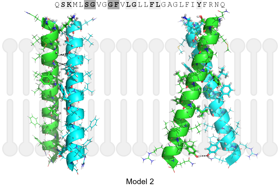
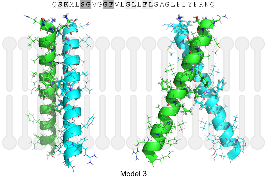
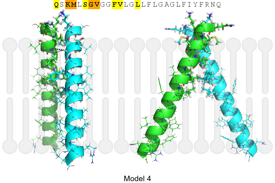
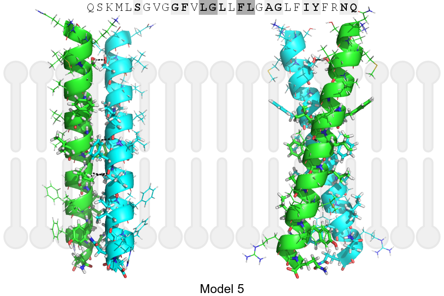
The sequence of 2B1A_HUMAN returned 5 potential models
The positions at the dimer's interface are highlighted in yellow. The position at the crossing point is highlighted in orange.
| # | Sequence and interface | d | θ | Z' | ω' | Score |
|---|---|---|---|---|---|---|
| 230 240 250 + | + | + | |
(Å) | (°) | (Å) | (°) | ||
| Model 1 | QSKMLSGVGGFVLGLLFLGAGLFIYFRNQ | 7.2 | -20.0° | 0.0 | 50.0 | -34.5 |
| Model 2 | QSKMLSGVGGFVLGLLFLGAGLFIYFRNQ | 7.1 | -36.1° | 2.4 | 59.2 | -20.9 |
| Model 3 | QSKMLSGVGGFVLGLLFLGAGLFIYFRNQ | 6.9 | -48.8° | 3.9 | 38.4 | -18.3 |
| Model 4 | QSKMLSGVGGFVLGLLFLGAGLFIYFRNQ | 7.2 | -51.0° | 4.3 | 40.0 | -9.7 |
| Model 5 | QSKMLSGVGGFVLGLLFLGAGLFIYFRNQ | 7.2 | -32.1° | 3.5 | 37.9 | -5.5 |
Click on the model number to visualize its details below.
MODEL 1 DETAILS
Precited structure:
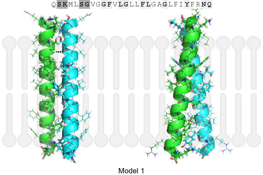
Click on the image to enlarge
Download model 1 structure: PDB, PyMOL session
List of hydrogen bonds:
| Donor | Acceptor | Distance |
|---|---|---|
| B, 231, GLY, HA2 | A, 230, SER, O | 3.12Å |
| B, 245, GLY, HA1 | A, 249, TYR, OH | 3.37Å |
| B, 246, LEU, HA | A, 249, TYR, OH | 2.85Å |
| A, 231, GLY, HA2 | B, 230, SER, O | 3.12Å |