UniProt data
UniProt name: ERBB4_HUMAN (Q15303)
Gene name: ERBB4; Synonyms=HER4
Organism: Homo sapiens (Human).
Function: Tyrosine-protein kinase that plays an essential role as cell surface receptor for neuregulins and EGF family members and regulates development of the heart, the central nervous system and the mammary gland, gene transcription, cell proliferation, differentiation, migration and apoptosis. Required for normal cardiac muscle differentiation during embryonic development, and for postnatal cardiomyocyte proliferation. Required for normal development of the embryonic central nervous system, especially for normal neural crest cell migration and normal axon guidance. Required for mammary gland differentiation, induction of milk proteins and lactation. Acts as cell-surface receptor for the neuregulins NRG1, NRG2, NRG3 and NRG4 and the EGF family members BTC, EREG and HBEGF. Ligand binding triggers receptor dimerization and autophosphorylation at specific tyrosine residues that then serve as binding sites for scaffold proteins and effectors. Ligand specificity and signaling is modulated by alternative splicing, proteolytic processing, and by the formation of heterodimers with other ERBB family members, thereby creating multiple combinations of intracellular phosphotyrosines that trigger ligand- and context-specific cellular responses. Mediates phosphorylation of SHC1 and activation of the MAP kinases MAPK1/ERK2 and MAPK3/ERK1. Isoform JM-A CYT-1 and isoform JM-B CYT-1 phosphorylate PIK3R1, leading to the activation of phosphatidylinositol 3-kinase and AKT1 and protect cells against apoptosis. Isoform JM-A CYT-1 and isoform JM-B CYT-1 mediate reorganization of the actin cytoskeleton and promote cell migration in response to NRG1. Isoform JM-A CYT-2 and isoform JM-B CYT-2 lack the phosphotyrosine that mediates interaction with PIK3R1, and hence do not phosphorylate PIK3R1, do not protect cells against apoptosis, and do not promote reorganization of the actin cytoskeleton and cell migration. Proteolytic processing of isoform JM-A CYT-1 and isoform JM-A CYT-2 gives rise to the corresponding soluble intracellular domains (4ICD) that translocate to the nucleus, promote nuclear import of STAT5A, activation of STAT5A, mammary epithelium differentiation, cell proliferation and activation of gene expression. The ERBB4 soluble intracellular domains (4ICD) colocalize with STAT5A at the CSN2 promoter to regulate transcription of milk proteins during lactation. The ERBB4 soluble intracellular domains can also translocate to mitochondria and promote apoptosis. {ECO:0000269|PubMed:10348342, ECO:0000269|PubMed:10353604, ECO:0000269|PubMed:10358079, ECO:0000269|PubMed:10722704, ECO:0000269|PubMed:10867024, ECO:0000269|PubMed:11178955, ECO:0000269|PubMed:11390655, ECO:0000269|PubMed:12807903, ECO:0000269|PubMed:15534001, ECO:0000269|PubMed:15746097, ECO:0000269|PubMed:16251361, ECO:0000269|PubMed:16778220, ECO:0000269|PubMed:16837552, ECO:0000269|PubMed:17486069, ECO:0000269|PubMed:17638867, ECO:0000269|PubMed:19098003, ECO:0000269|PubMed:20858735, ECO:0000269|PubMed:8383326, ECO:0000269|PubMed:8617750, ECO:0000269|PubMed:9135143, ECO:0000269|PubMed:9168115, ECO:0000269|PubMed:9334263}.
Subcellular location: ERBB4 intracellular domain: Nucleus {ECO:0000269|PubMed:17486069}. Mitochondrion {ECO:0000269|PubMed:17486069}. Note=Following proteolytical processing E4ICD (E4ICD1 or E4ICD2 generated from the respective isoforms) is translocated to the nucleus. Significantly more E4ICD2 than E4ICD1 is found in the nucleus. E4ICD2 colocalizes with YAP1 in the nucleus.
Transmembrane domains:
- 652-675, 24 AA, Helical. {ECO:0000255}.
Sequence: 1308 AA
CATM prediction
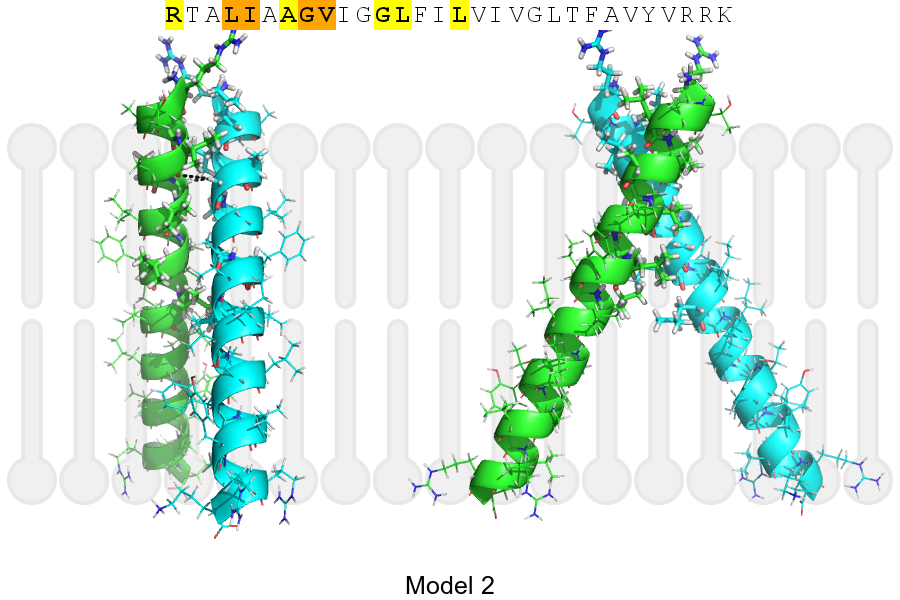
The sequence of ERBB4_HUMAN returned 2 potential models
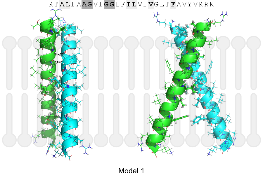
The positions at the dimer's interface are highlighted in yellow. The position at the crossing point is highlighted in orange.
| # | Sequence and interface | d | θ | Z' | ω' | Score |
|---|---|---|---|---|---|---|
| 650 660 670 | + | + | + |
(Å) | (°) | (Å) | (°) | ||
| Model 1 | RTALIAAGVIGGLFILVIVGLTFAVYVRRK | 6.7 | -46.0° | 3.6 | 37.0 | -18.8 |
| Model 2 | RTALIAAGVIGGLFILVIVGLTFAVYVRRK | 7.2 | -53.0° | 1.9 | 45.0 | -4.7 |
| Prolines* | RTPLIAAGVIGGLFILVIVGLTFAVYVRRK |
* Note: this sequence contains Pro residues. CATM substituted them with Ala during the modeling. The positions are marked.
Click on the model number to visualize its details below.
MODEL 1 DETAILS
Precited structure:
Click on the image to enlarge
Download model 1 structure: PDB, PyMOL session
List of hydrogen bonds:
| Donor | Acceptor | Distance |
|---|---|---|
| B, 656, GLY, HA2 | A, 655, ALA, O | 2.75Å |
| B, 659, GLY, HA1 | A, 656, GLY, O | 2.80Å |
| A, 656, GLY, HA2 | B, 655, ALA, O | 2.75Å |
| A, 659, GLY, HA1 | B, 656, GLY, O | 2.80Å |