UniProt data
UniProt name: FKBP8_HUMAN (Q14318)
Gene name: FKBP8; Synonyms=FKBP38
Organism: Homo sapiens (Human).
Function: Constitutively inactive PPiase, which becomes active when bound to calmodulin and calcium. Seems to act as a chaperone for BCL2, targets it to the mitochondria and modulates its phosphorylation state. The BCL2/FKBP8/calmodulin/calcium complex probably interferes with the binding of BCL2 to its targets. The active form of FKBP8 may therefore play a role in the regulation of apoptosis. {ECO:0000269|PubMed:12510191, ECO:0000269|PubMed:15757646, ECO:0000269|PubMed:16176796}.
Subcellular location: Isoform 3: Mitochondrion membrane {ECO:0000269|PubMed:18385096}; Single-pass membrane protein; Cytoplasmic side {ECO:0000305}.
Transmembrane domains:
- 390-410, 21 AA, Helical. {ECO:0000255}.
Sequence: 412 AA
CATM prediction
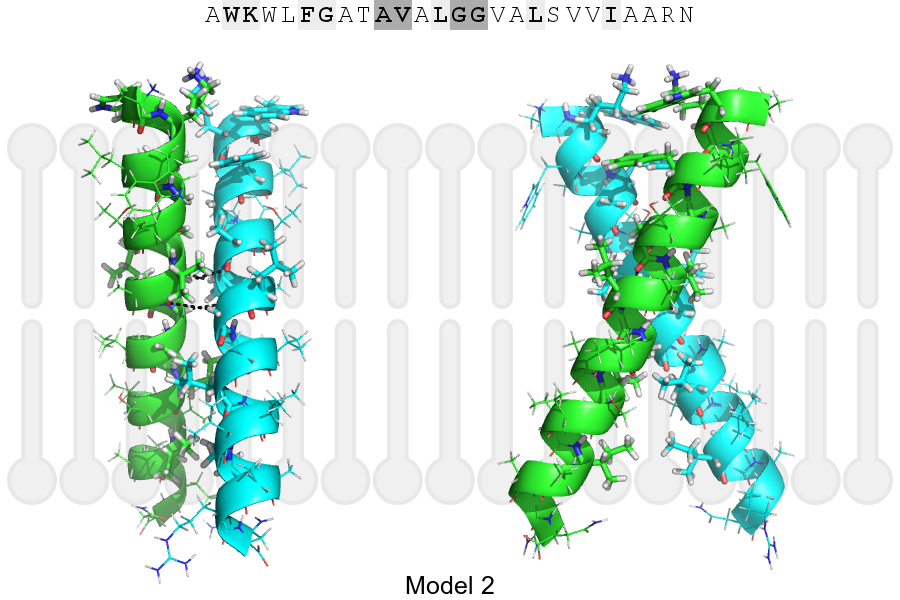
The sequence of FKBP8_HUMAN returned 2 potential models
The positions at the dimer's interface are highlighted in yellow. The position at the crossing point is highlighted in orange.
| # | Sequence and interface | d | θ | Z' | ω' | Score |
|---|---|---|---|---|---|---|
| 390 400 410 | + | + | |
(Å) | (°) | (Å) | (°) | ||
| Model 1 | AWKWLFGATAVALGGVALSVVIAARN | 7.3 | -44.0° | 1.8 | 45.0 | -7.0 |
| Model 2 | AWKWLFGATAVALGGVALSVVIAARN | 7.3 | -53.9° | 3.6 | 49.2 | -2.6 |
| Prolines* | PWKWLFGATAVALGGVALSVVIAARN |
* Note: this sequence contains Pro residues. CATM substituted them with Ala during the modeling. The positions are marked.
Click on the model number to visualize its details below.
MODEL 1 DETAILS
Precited structure:
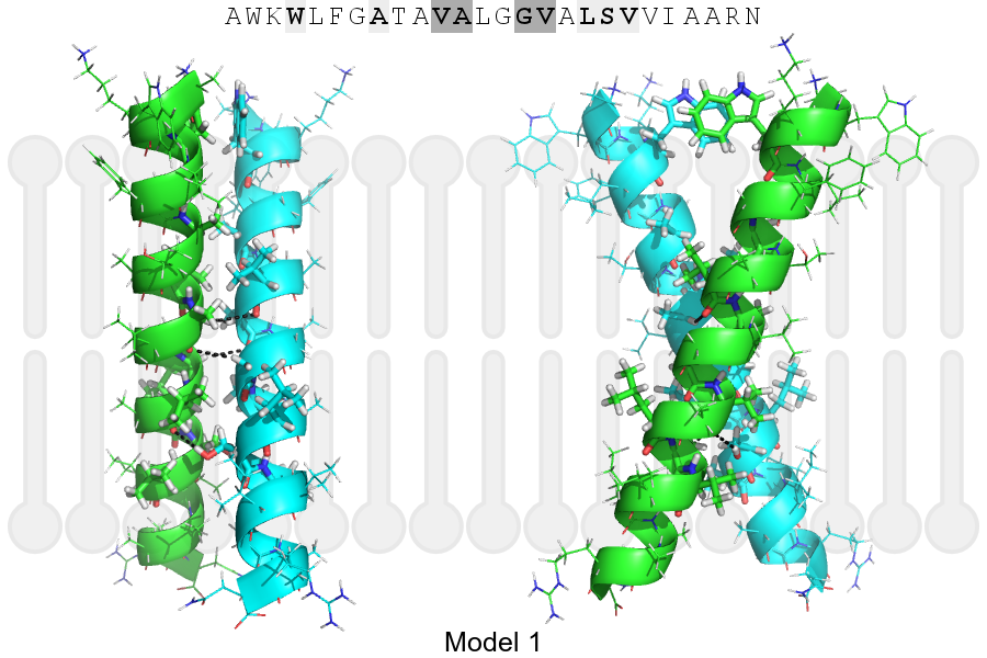
Click on the image to enlarge
Download model 1 structure: PDB, PyMOL session
List of hydrogen bonds:
| Donor | Acceptor | Distance |
|---|---|---|
| B, 398, ALA, HA | A, 397, VAL, O | 3.47Å |
| B, 401, GLY, HA1 | A, 398, ALA, O | 3.26Å |
| B, 405, SER, HG1 | A, 402, VAL, O | 2.57Å |
| A, 398, ALA, HA | B, 397, VAL, O | 3.47Å |
| A, 401, GLY, HA1 | B, 398, ALA, O | 3.26Å |
| A, 405, SER, HG1 | B, 402, VAL, O | 2.57Å |