

UniProt data
UniProt name: DAG1_HUMAN (Q14118)
Gene name: DAG1
Organism: Homo sapiens (Human).
Function: (Microbial infection) Alpha-dystroglycan acts as a receptor for lassa virus and lymphocytic choriomeningitis virus glycoprotein and class C new-world arenaviruses (PubMed:16254364, PubMed:19324387, PubMed:17360738). Alpha-dystroglycan acts as a Schwann cell receptor for Mycobacterium leprae, the causative organism of leprosy, but only in the presence of the G-domain of LAMA2 (PubMed:9851927). {ECO:0000269|PubMed:16254364, ECO:0000269|PubMed:17360738, ECO:0000269|PubMed:19324387, ECO:0000269|PubMed:9851927}.
Subcellular location: Beta-dystroglycan: Cell membrane; Single- pass type I membrane protein. Cytoplasm, cytoskeleton. Nucleus, nucleoplasm. Cell membrane, sarcolemma {ECO:0000250}. Cell junction, synapse, postsynaptic cell membrane {ECO:0000250}. Note=The monomeric form translocates to the nucleus via the action of importins and depends on RAN. Nuclear transport is inhibited by Tyr-892 phosphorylation. In skeletal muscle, this phosphorylated form locates to a vesicular internal membrane compartment. In muscle cells, sarcolemma localization requires the presence of ANK2, while localization to costameres requires the presence of ANK3. Localizes to neuromuscular junctions (NMJs) in the presence of ANK2 (By similarity). In peripheral nerves, localizes to the Schwann cell membrane. Colocalizes with ERM proteins in Schwann- cell microvilli. {ECO:0000250}.
Transmembrane domains:
- 750-775, 26 AA, Helical. {ECO:0000255}.
Sequence: 895 AA
CATM prediction
The sequence of DAG1_HUMAN returned 1 potential models
The positions at the dimer's interface are highlighted in yellow. The position at the crossing point is highlighted in orange.
| # | Sequence and interface | d | θ | Z' | ω' | Score |
|---|---|---|---|---|---|---|
| 750 760 770 | + | + | + |
(Å) | (°) | (Å) | (°) | ||
| Model 1 | DDVYLHTVIAAVVVAAILLIAGIIAMICYRKK | 7.3 | -55.0° | 2.4 | 48.0 | -10.9 |
| Prolines* | DDVYLHTVIPAVVVAAILLIAGIIAMICYRKK |
* Note: this sequence contains Pro residues. CATM substituted them with Ala during the modeling. The positions are marked.
Click on the model number to visualize its details below.
MODEL 1 DETAILS
Precited structure:
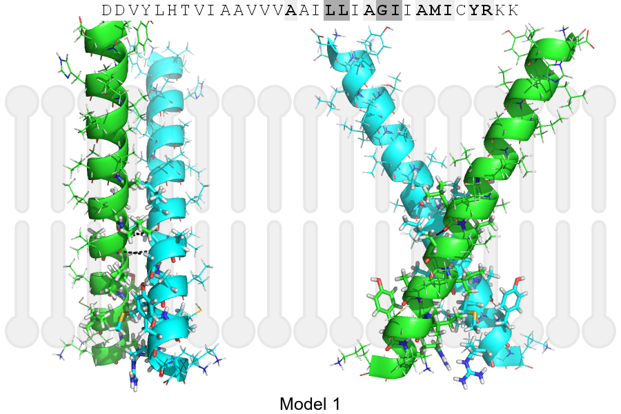
Click on the image to enlarge
Download model 1 structure: PDB, PyMOL session
List of hydrogen bonds:
| Donor | Acceptor | Distance |
|---|---|---|
| B, 765, LEU, HA | A, 764, LEU, O | 3.48Å |
| B, 768, GLY, HA1 | A, 765, LEU, O | 3.23Å |
| A, 765, LEU, HA | B, 764, LEU, O | 3.48Å |
| A, 768, GLY, HA1 | B, 765, LEU, O | 3.23Å |