UniProt data
UniProt name: STIM1_HUMAN (Q13586)
Gene name: STIM1; Synonyms=GOK {ECO:0000303|PubMed:9377559}
Organism: Homo sapiens (Human).
Function: Plays a role in mediating store-operated Ca(2+) entry (SOCE), a Ca(2+) influx following depletion of intracellular Ca(2+) stores (PubMed:15866891, PubMed:16005298, PubMed:16208375, PubMed:16537481, PubMed:16733527, PubMed:16766533, PubMed:16807233, PubMed:18854159, PubMed:19249086, PubMed:22464749, PubMed:24069340, PubMed:24351972, PubMed:24591628, PubMed:26322679, PubMed:25326555). Acts as Ca(2+) sensor in the endoplasmic reticulum via its EF-hand domain. Upon Ca(2+) depletion, translocates from the endoplasmic reticulum to the plasma membrane where it activates the Ca(2+) release- activated Ca(2+) (CRAC) channel subunit ORAI1 (PubMed:16208375, PubMed:16537481). Involved in enamel formation (PubMed:24621671). Activated following interaction with TMEM110/STIMATE, leading to promote STIM1 conformational switch (PubMed:26322679). {ECO:0000269|PubMed:15866891, ECO:0000269|PubMed:16005298, ECO:0000269|PubMed:16208375, ECO:0000269|PubMed:16537481, ECO:0000269|PubMed:16733527, ECO:0000269|PubMed:16766533, ECO:0000269|PubMed:16807233, ECO:0000269|PubMed:18854159, ECO:0000269|PubMed:19249086, ECO:0000269|PubMed:22464749, ECO:0000269|PubMed:24069340, ECO:0000269|PubMed:24351972, ECO:0000269|PubMed:24591628, ECO:0000269|PubMed:24621671, ECO:0000269|PubMed:25326555, ECO:0000269|PubMed:26322679}.
Subcellular location: Cell membrane; Single-pass type I membrane protein {ECO:0000269|PubMed:11004585, ECO:0000269|PubMed:16005298, ECO:0000269|PubMed:16208375, ECO:0000269|PubMed:18854159, ECO:0000269|PubMed:19249086}. Endoplasmic reticulum membrane; Single-pass type I membrane protein {ECO:0000269|PubMed:16005298, ECO:0000269|PubMed:16208375, ECO:0000269|PubMed:18854159, ECO:0000269|PubMed:19249086, ECO:0000269|PubMed:26322679}. Cytoplasm, cytoskeleton {ECO:0000269|PubMed:19632184}. Sarcoplasmic reticulum {ECO:0000269|PubMed:25326555}. Note=Translocates from the endoplasmic reticulum to the cell membrane in response to a depletion of intracellular calcium and is detected at punctae corresponding to junctions between the endoplasmic reticulum and the cell membrane (PubMed:19249086, PubMed:16005298, PubMed:16208375, PubMed:18854159). Associated with the microtubule network at the growing distal tip of microtubules (PubMed:19632184). {ECO:0000269|PubMed:16005298, ECO:0000269|PubMed:16208375, ECO:0000269|PubMed:18854159, ECO:0000269|PubMed:19249086, ECO:0000269|PubMed:19632184}.
Transmembrane domains:
- 214-234, 21 AA, Helical. {ECO:0000255}.
Sequence: 685 AA
CATM prediction
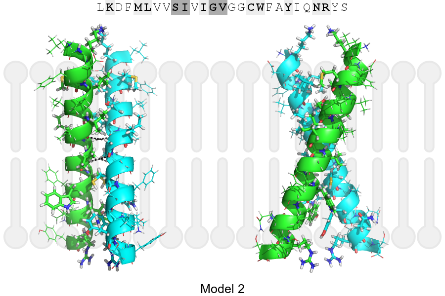
The sequence of STIM1_HUMAN returned 2 potential models
The positions at the dimer's interface are highlighted in yellow. The position at the crossing point is highlighted in orange.
| # | Sequence and interface | d | θ | Z' | ω' | Score |
|---|---|---|---|---|---|---|
| 220 230 + | + | + |
(Å) | (°) | (Å) | (°) | ||
| Model 1 | LKDFMLVVSIVIGVGGCWFAYIQNRYS | 7.3 | -37.8° | 3.1 | 35.1 | -28.2 |
| Model 2 | LKDFMLVVSIVIGVGGCWFAYIQNRYS | 7.3 | -46.9° | 1.4 | 45.1 | -7.7 |
Click on the model number to visualize its details below.
MODEL 1 DETAILS
Precited structure:
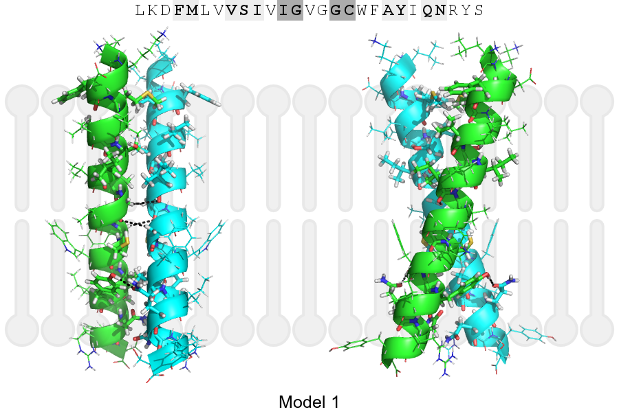
Click on the image to enlarge
Download model 1 structure: PDB, PyMOL session
List of hydrogen bonds:
| Donor | Acceptor | Distance |
|---|---|---|
| B, 223, GLY, HA2 | A, 222, ILE, O | 3.41Å |
| B, 226, GLY, HA1 | A, 223, GLY, O | 3.32Å |
| B, 231, TYR, HH | A, 233, GLN, OE1 | 2.27Å |
| A, 223, GLY, HA2 | B, 222, ILE, O | 3.41Å |
| A, 226, GLY, HA1 | B, 223, GLY, O | 3.32Å |
| A, 231, TYR, HH | B, 233, GLN, OE1 | 2.27Å |