

UniProt data
UniProt name: NRG1_HUMAN (Q02297)
Gene name: NRG1; Synonyms=GGF, HGL, HRGA, NDF, SMDF
Organism: Homo sapiens (Human).
Function: Direct ligand for ERBB3 and ERBB4 tyrosine kinase receptors. Concomitantly recruits ERBB1 and ERBB2 coreceptors, resulting in ligand-stimulated tyrosine phosphorylation and activation of the ERBB receptors. The multiple isoforms perform diverse functions such as inducing growth and differentiation of epithelial, glial, neuronal, and skeletal muscle cells; inducing expression of acetylcholine receptor in synaptic vesicles during the formation of the neuromuscular junction; stimulating lobuloalveolar budding and milk production in the mammary gland and inducing differentiation of mammary tumor cells; stimulating Schwann cell proliferation; implication in the development of the myocardium such as trabeculation of the developing heart. Isoform 10 may play a role in motor and sensory neuron development. Binds to ERBB4 (PubMed:10867024, PubMed:7902537). Binds to ERBB3 (PubMed:20682778). Acts as a ligand for integrins and binds (via EGF domain) to integrins ITGAV:ITGB3 or ITGA6:ITGB4. Its binding to integrins and subsequent ternary complex formation with integrins and ERRB3 are essential for NRG1-ERBB signaling. Induces the phosphorylation and activation of MAPK3/ERK1, MAPK1/ERK2 and AKT1 (PubMed:20682778). Ligand-dependent ERBB4 endocytosis is essential for the NRG1-mediated activation of these kinases in neurons (By similarity). {ECO:0000250|UniProtKB:P43322, ECO:0000269|PubMed:10867024, ECO:0000269|PubMed:1348215, ECO:0000269|PubMed:20682778, ECO:0000269|PubMed:7902537}.
Subcellular location: Isoform 10: Membrane; Single-pass type I membrane protein. Note=May possess an internal uncleaved signal sequence.
Transmembrane domains:
- 243-265, 23 AA, Helical; Note=Internal signal sequence. {ECO:0000255}.
Sequence: 640 AA
CATM prediction
The sequence of NRG1_HUMAN returned 1 potential models
The positions at the dimer's interface are highlighted in yellow. The position at the crossing point is highlighted in orange.
| # | Sequence and interface | d | θ | Z' | ω' | Score |
|---|---|---|---|---|---|---|
| 240 250 260 | + | + | + |
(Å) | (°) | (Å) | (°) | ||
| Model 1 | QKRVLTITGICIALLVVGIMCVVAYCKTK | 7.3 | -47.1° | 1.3 | 45.0 | -15.6 |
Click on the model number to visualize its details below.
MODEL 1 DETAILS
Precited structure:
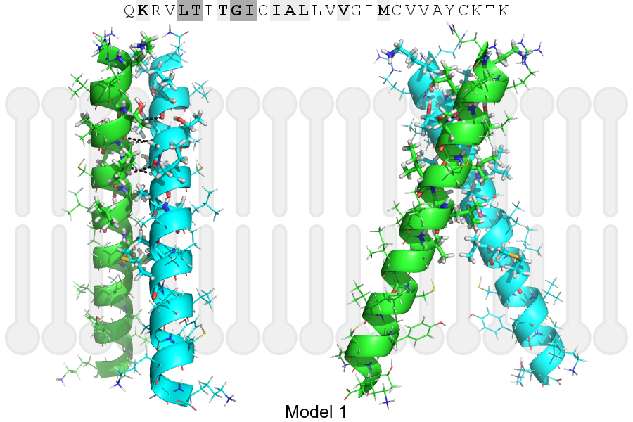
Click on the image to enlarge
Download model 1 structure: PDB, PyMOL session
List of hydrogen bonds:
| Donor | Acceptor | Distance |
|---|---|---|
| B, 245, THR, HA | A, 244, LEU, O | 3.48Å |
| B, 248, GLY, HA1 | A, 245, THR, O | 3.09Å |
| B, 249, ILE, HA | A, 248, GLY, O | 3.26Å |
| A, 245, THR, HA | B, 244, LEU, O | 3.48Å |
| A, 248, GLY, HA1 | B, 245, THR, O | 3.09Å |
| A, 249, ILE, HA | B, 248, GLY, O | 3.26Å |