

UniProt data
UniProt name: EFNB1_HUMAN (P98172)
Gene name: EFNB1; Synonyms=EFL3, EPLG2, LERK2
Organism: Homo sapiens (Human).
Function: Cell surface transmembrane ligand for Eph receptors, a family of receptor tyrosine kinases which are crucial for migration, repulsion and adhesion during neuronal, vascular and epithelial development. Binds promiscuously Eph receptors residing on adjacent cells, leading to contact-dependent bidirectional signaling into neighboring cells. The signaling pathway downstream of the receptor is referred to as forward signaling while the signaling pathway downstream of the ephrin ligand is referred to as reverse signaling. Binds to the receptor tyrosine kinases EPHB3 (preferred), EPHB1 and EPHA1. Binds to, and induce the collapse of, commissural axons/growth cones in vitro. May play a role in constraining the orientation of longitudinally projecting axons (By similarity). {ECO:0000250}.
Subcellular location: Membrane; Single-pass type I membrane protein.
Transmembrane domains:
- 238-258, 21 AA, Helical. {ECO:0000255}.
Sequence: 346 AA
CATM prediction
The sequence of EFNB1_HUMAN returned 1 potential models
The positions at the dimer's interface are highlighted in yellow. The position at the crossing point is highlighted in orange.
| # | Sequence and interface | d | θ | Z' | ω' | Score |
|---|---|---|---|---|---|---|
| 240 250 26 + | + | + | |
(Å) | (°) | (Å) | (°) | ||
| Model 1 | NSKVALFAAVGAGCVIFLLIIIFLTVL | 7.0 | -50.9° | 2.4 | 45.1 | -37.4 |
Click on the model number to visualize its details below.
MODEL 1 DETAILS
Precited structure:
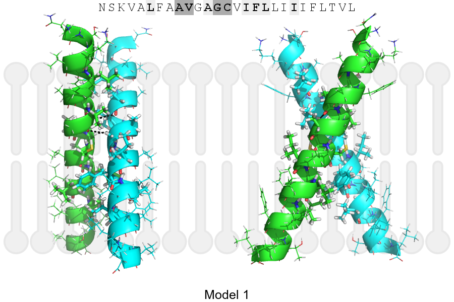
Click on the image to enlarge
Download model 1 structure: PDB, PyMOL session
List of hydrogen bonds:
| Donor | Acceptor | Distance |
|---|---|---|
| B, 244, VAL, HA | A, 243, ALA, O | 3.18Å |
| B, 247, GLY, HA1 | A, 244, VAL, O | 2.98Å |
| A, 244, VAL, HA | B, 243, ALA, O | 3.18Å |
| A, 247, GLY, HA1 | B, 244, VAL, O | 2.98Å |