

UniProt data
UniProt name: X3CL1_HUMAN (P78423)
Gene name: CX3CL1; Synonyms=FKN, NTT, SCYD1; ORFNames=A-152E5.2
Organism: Homo sapiens (Human).
Function: Acts as a ligand for both CX3CL1 and integrins. Binds to CX3CR1 (PubMed:23125415, PubMed:9931005, PubMed:21829356). Binds to integrins ITGAV:ITGB3 and ITGA4:ITGB1. Can activate integrins in both a CX3CR1-dependent and CX3CR1-independent manner. In the presence of CX3CR1, activates integrins by binding to the classical ligand-binding site (site 1) in integrins. In the absence of CX3CR1, binds to a second site (site 2) in integrins which is distinct from site 1 and enhances the binding of other integrin ligands to site 1 (PubMed:23125415, PubMed:24789099). The soluble form is chemotactic for T-cells and monocytes and not for neutrophils. The membrane-bound form promotes adhesion of those leukocytes to endothelial cells. May play a role in regulating leukocyte adhesion and migration processes at the endothelium (PubMed:9024663, PubMed:9177350). {ECO:0000269|PubMed:21829356, ECO:0000269|PubMed:23125415, ECO:0000269|PubMed:24789099, ECO:0000269|PubMed:9024663, ECO:0000269|PubMed:9177350, ECO:0000269|PubMed:9931005}.
Subcellular location: Processed fractalkine: Secreted {ECO:0000269|PubMed:9024663}.
Transmembrane domains:
- 342-362, 21 AA, Helical. {ECO:0000255}.
Sequence: 397 AA
CATM prediction
The sequence of X3CL1_HUMAN returned 1 potential models
The positions at the dimer's interface are highlighted in yellow. The position at the crossing point is highlighted in orange.
| # | Sequence and interface | d | θ | Z' | ω' | Score |
|---|---|---|---|---|---|---|
| 340 350 360 | + | + | + |
(Å) | (°) | (Å) | (°) | ||
| Model 1 | RRQAVGLLAFLGLLFCLGVAMFTYQSL | 7.3 | -55.0° | 4.2 | 38.0 | -1.0 |
Click on the model number to visualize its details below.
MODEL 1 DETAILS
Precited structure:
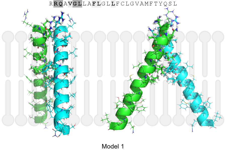
Click on the image to enlarge
Download model 1 structure: PDB, PyMOL session