

UniProt data
UniProt name: SHPS1_HUMAN (P78324)
Gene name: SIRPA; Synonyms=BIT, MFR, MYD1, PTPNS1, SHPS1, SIRP
Organism: Homo sapiens (Human).
Function: Immunoglobulin-like cell surface receptor for CD47. Acts as docking protein and induces translocation of PTPN6, PTPN11 and other binding partners from the cytosol to the plasma membrane. Supports adhesion of cerebellar neurons, neurite outgrowth and glial cell attachment. May play a key role in intracellular signaling during synaptogenesis and in synaptic function (By similarity). Involved in the negative regulation of receptor tyrosine kinase-coupled cellular responses induced by cell adhesion, growth factors or insulin. Mediates negative regulation of phagocytosis, mast cell activation and dendritic cell activation. CD47 binding prevents maturation of immature dendritic cells and inhibits cytokine production by mature dendritic cells. {ECO:0000250, ECO:0000269|PubMed:10469599, ECO:0000269|PubMed:11509594}.
Subcellular location: Membrane; Single-pass type I membrane protein.
Transmembrane domains:
- 374-394, 21 AA, Helical. {ECO:0000255}.
Sequence: 504 AA
CATM prediction
The sequence of SHPS1_HUMAN returned 1 potential models
The positions at the dimer's interface are highlighted in yellow. The position at the crossing point is highlighted in orange.
| # | Sequence and interface | d | θ | Z' | ω' | Score |
|---|---|---|---|---|---|---|
| 380 390 + | + | + |
(Å) | (°) | (Å) | (°) | ||
| Model 1 | NIYIVVGVVCTLLVALLMAALYLVRIR | 7.3 | -50.5° | 3.3 | 48.0 | -10.0 |
Click on the model number to visualize its details below.
MODEL 1 DETAILS
Precited structure:
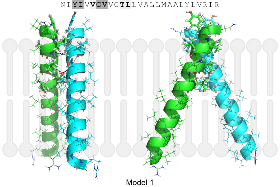
Click on the image to enlarge
Download model 1 structure: PDB, PyMOL session
List of hydrogen bonds:
| Donor | Acceptor | Distance |
|---|---|---|
| B, 377, GLY, HA1 | A, 374, ILE, O | 3.28Å |
| B, 378, VAL, HA | A, 381, THR, OG1 | 2.83Å |
| A, 377, GLY, HA1 | B, 374, ILE, O | 3.28Å |
| A, 378, VAL, HA | B, 381, THR, OG1 | 2.83Å |