UniProt data
UniProt name: ATPK_HUMAN (P56134)
Gene name: ATP5J2; Synonyms=ATP5JL
Organism: Homo sapiens (Human).
Function: Mitochondrial membrane ATP synthase (F(1)F(0) ATP synthase or Complex V) produces ATP from ADP in the presence of a proton gradient across the membrane which is generated by electron transport complexes of the respiratory chain. F-type ATPases consist of two structural domains, F(1) - containing the extramembraneous catalytic core and F(0) - containing the membrane proton channel, linked together by a central stalk and a peripheral stalk. During catalysis, ATP synthesis in the catalytic domain of F(1) is coupled via a rotary mechanism of the central stalk subunits to proton translocation. Part of the complex F(0) domain. Minor subunit located with subunit a in the membrane.
Subcellular location: Mitochondrion. Mitochondrion inner membrane {ECO:0000305}; Single-pass membrane protein {ECO:0000305}.
Transmembrane domains:
- 63-82, 20 AA, Helical. {ECO:0000255}.
Sequence: 94 AA
CATM prediction
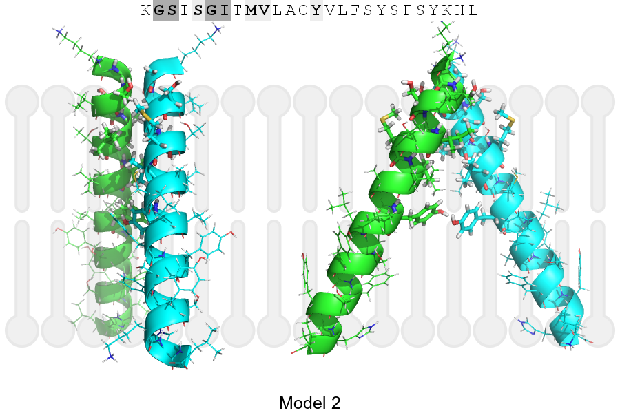
The sequence of ATPK_HUMAN returned 2 potential models
The positions at the dimer's interface are highlighted in yellow. The position at the crossing point is highlighted in orange.
| # | Sequence and interface | d | θ | Z' | ω' | Score |
|---|---|---|---|---|---|---|
| 60 70 80 | + | + | + |
(Å) | (°) | (Å) | (°) | ||
| Model 1 | KGSISGITMVLACYVLFSYSFSYKHL | 7.0 | -43.0° | 5.5 | 34.0 | -9.3 |
| Model 2 | KGSISGITMVLACYVLFSYSFSYKHL | 6.5 | -55.0° | 5.8 | 47.0 | -3.4 |
Click on the model number to visualize its details below.
MODEL 1 DETAILS
Precited structure:
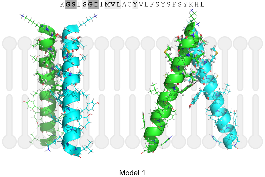
Click on the image to enlarge
Download model 1 structure: PDB, PyMOL session
List of hydrogen bonds:
| Donor | Acceptor | Distance |
|---|---|---|
| B, 70, LEU, HA | A, 73, TYR, OH | 2.65Å |