

UniProt data
UniProt name: IOD3_HUMAN (P55073)
Gene name: DIO3; Synonyms=ITDI3, TXDI3
Organism: Homo sapiens (Human).
Function: Responsible for the deiodination of T4 (3,5,3',5'- tetraiodothyronine) into RT3 (3,3',5'-triiodothyronine) and of T3 (3,5,3'-triiodothyronine) into T2 (3,3'-diiodothyronine). RT3 and T2 are inactive metabolites. May play a role in preventing premature exposure of developing fetal tissues to adult levels of thyroid hormones. Can regulate circulating fetal thyroid hormone concentrations throughout gestation. Essential role for regulation of thyroid hormone inactivation during embryological development. {ECO:0000269|PubMed:7593630}.
Subcellular location: Cell membrane {ECO:0000269|PubMed:12419801}; Single-pass type II membrane protein {ECO:0000269|PubMed:12419801}. Endosome membrane {ECO:0000269|PubMed:12419801}; Single-pass type II membrane protein {ECO:0000269|PubMed:12419801}.
Transmembrane domains:
- 45-67, 23 AA, Helical; Signal-anchor for type II membrane protein. {ECO:0000255}.
Sequence: 304 AA
CATM prediction
The sequence of IOD3_HUMAN returned 1 potential models
The positions at the dimer's interface are highlighted in yellow. The position at the crossing point is highlighted in orange.
| # | Sequence and interface | d | θ | Z' | ω' | Score |
|---|---|---|---|---|---|---|
| 50 60 7 + | + | + | |
(Å) | (°) | (Å) | (°) | ||
| Model 1 | TASCLVLFARFLGTAFMLWLLDFLCIRKH | 6.8 | -50.0° | 2.9 | 46.9 | -24.6 |
| Prolines* | TASCLVLFPRFLGTAFMLWLLDFLCIRKH |
* Note: this sequence contains Pro residues. CATM substituted them with Ala during the modeling. The positions are marked.
Click on the model number to visualize its details below.
MODEL 1 DETAILS
Precited structure:
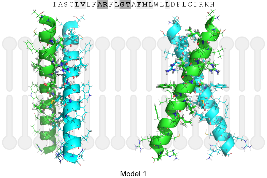
Click on the image to enlarge
Download model 1 structure: PDB, PyMOL session
List of hydrogen bonds:
| Donor | Acceptor | Distance |
|---|---|---|
| B, 51, ARG, HA | A, 50, ALA, O | 2.87Å |
| B, 54, GLY, HA1 | A, 51, ARG, O | 2.87Å |
| A, 51, ARG, HA | B, 50, ALA, O | 2.87Å |
| A, 54, GLY, HA1 | B, 51, ARG, O | 2.87Å |