

UniProt data
UniProt name: EPHB1_HUMAN (P54762)
Gene name: EPHB1; Synonyms=ELK, EPHT2, HEK6, NET
Organism: Homo sapiens (Human).
Function: Receptor tyrosine kinase which binds promiscuously transmembrane ephrin-B family ligands residing on adjacent cells, leading to contact-dependent bidirectional signaling into neighboring cells. The signaling pathway downstream of the receptor is referred to as forward signaling while the signaling pathway downstream of the ephrin ligand is referred to as reverse signaling. Cognate/functional ephrin ligands for this receptor include EFNB1, EFNB2 and EFNB3. During nervous system development, regulates retinal axon guidance redirecting ipsilaterally ventrotemporal retinal ganglion cells axons at the optic chiasm midline. This probably requires repulsive interaction with EFNB2. In the adult nervous system together with EFNB3, regulates chemotaxis, proliferation and polarity of the hippocampus neural progenitors. In addition to its role in axon guidance plays also an important redundant role with other ephrin-B receptors in development and maturation of dendritic spines and synapse formation. May also regulate angiogenesis. More generally, may play a role in targeted cell migration and adhesion. Upon activation by EFNB1 and probably other ephrin-B ligands activates the MAPK/ERK and the JNK signaling cascades to regulate cell migration and adhesion respectively. {ECO:0000269|PubMed:12223469, ECO:0000269|PubMed:12925710, ECO:0000269|PubMed:18034775, ECO:0000269|PubMed:9430661, ECO:0000269|PubMed:9499402}.
Subcellular location: Cell membrane {ECO:0000269|PubMed:18034775}; Single-pass type I membrane protein {ECO:0000269|PubMed:18034775}. Early endosome membrane {ECO:0000269|PubMed:18034775}. Cell projection, dendrite {ECO:0000250}.
Transmembrane domains:
- 541-563, 23 AA, Helical. {ECO:0000255}.
Sequence: 984 AA
CATM prediction
The sequence of EPHB1_HUMAN returned 1 potential models
The positions at the dimer's interface are highlighted in yellow. The position at the crossing point is highlighted in orange.
| # | Sequence and interface | d | θ | Z' | ω' | Score |
|---|---|---|---|---|---|---|
| 540 550 560 | + | + | + |
(Å) | (°) | (Å) | (°) | ||
| Model 1 | QLALIAGSAAAGVVFVVSLVAISIVCSRK | 6.9 | -42.9° | 2.4 | 43.9 | -17.0 |
| Prolines* | QLPLIAGSAAAGVVFVVSLVAISIVCSRK |
* Note: this sequence contains Pro residues. CATM substituted them with Ala during the modeling. The positions are marked.
Click on the model number to visualize its details below.
MODEL 1 DETAILS
Precited structure:
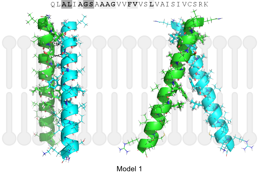
Click on the image to enlarge
Download model 1 structure: PDB, PyMOL session
List of hydrogen bonds:
| Donor | Acceptor | Distance |
|---|---|---|
| B, 544, GLY, HA1 | A, 541, LEU, O | 2.87Å |
| B, 545, SER, HA | A, 544, GLY, O | 3.34Å |
| A, 544, GLY, HA1 | B, 541, LEU, O | 2.87Å |
| A, 545, SER, HA | B, 544, GLY, O | 3.34Å |