UniProt data
UniProt name: EPHB3_HUMAN (P54753)
Gene name: EPHB3; Synonyms=ETK2, HEK2, TYRO6
Organism: Homo sapiens (Human).
Function: Receptor tyrosine kinase which binds promiscuously transmembrane ephrin-B family ligands residing on adjacent cells, leading to contact-dependent bidirectional signaling into neighboring cells. The signaling pathway downstream of the receptor is referred to as forward signaling while the signaling pathway downstream of the ephrin ligand is referred to as reverse signaling. Generally has an overlapping and redundant function with EPHB2. Like EPHB2, functions in axon guidance during development regulating for instance the neurons forming the corpus callosum and the anterior commissure, 2 major interhemispheric connections between the temporal lobes of the cerebral cortex. In addition to its role in axon guidance plays also an important redundant role with other ephrin-B receptors in development and maturation of dendritic spines and the formation of excitatory synapses. Controls other aspects of development through regulation of cell migration and positioning. This includes angiogenesis, palate development and thymic epithelium development for instance. Forward and reverse signaling through the EFNB2/EPHB3 complex also regulate migration and adhesion of cells that tubularize the urethra and septate the cloaca. Finally, plays an important role in intestinal epithelium differentiation segregating progenitor from differentiated cells in the crypt. {ECO:0000269|PubMed:15536074}.
Subcellular location: Cell membrane {ECO:0000269|PubMed:15536074}; Single-pass type I membrane protein {ECO:0000269|PubMed:15536074}. Cell projection, dendrite {ECO:0000250}.
Transmembrane domains:
- 560-580, 21 AA, Helical. {ECO:0000255}.
Sequence: 998 AA
CATM prediction
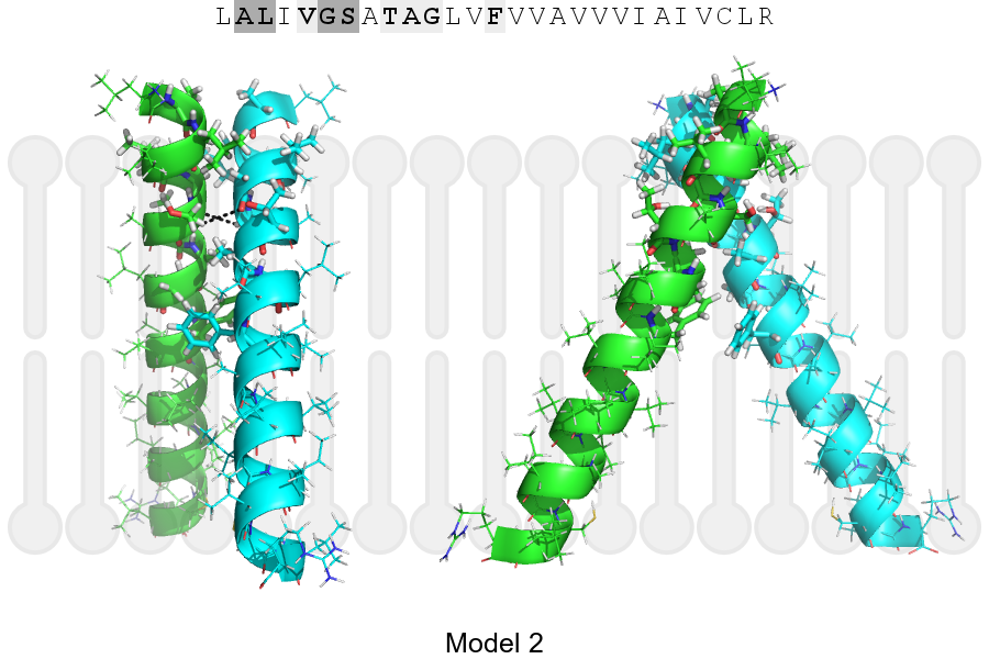
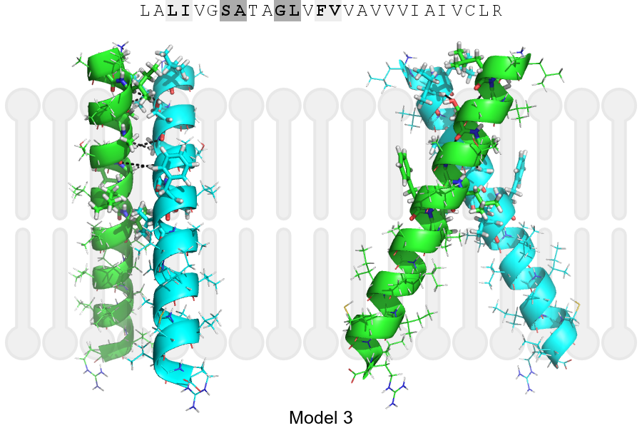
The sequence of EPHB3_HUMAN returned 3 potential models
The positions at the dimer's interface are highlighted in yellow. The position at the crossing point is highlighted in orange.
| # | Sequence and interface | d | θ | Z' | ω' | Score |
|---|---|---|---|---|---|---|
| 560 570 580 | + | + | |
(Å) | (°) | (Å) | (°) | ||
| Model 1 | LALIVGSATAGLVFVVAVVVIAIVCLR | 7.3 | -35.0° | 5.0 | 35.0 | -21.3 |
| Model 2 | LALIVGSATAGLVFVVAVVVIAIVCLR | 7.1 | -55.0° | 0.9 | 50.0 | -10.2 |
| Model 3 | LALIVGSATAGLVFVVAVVVIAIVCLR | 7.3 | -54.0° | 3.6 | 49.1 | -9.0 |
| Prolines* | LPLIVGSATAGLVFVVAVVVIAIVCLR |
* Note: this sequence contains Pro residues. CATM substituted them with Ala during the modeling. The positions are marked.
Click on the model number to visualize its details below.
MODEL 1 DETAILS
Precited structure:
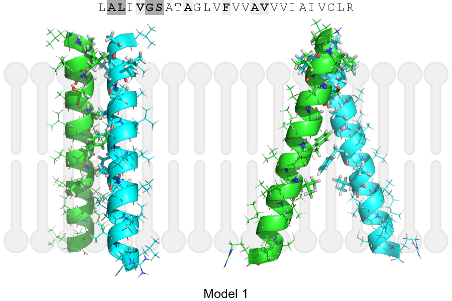
Click on the image to enlarge
Download model 1 structure: PDB, PyMOL session