

UniProt data
UniProt name: EMD_HUMAN (P50402)
Gene name: EMD; Synonyms=EDMD, STA
Organism: Homo sapiens (Human).
Function: Stabilizes and promotes the formation of a nuclear actin cortical network. Stimulates actin polymerization in vitro by binding and stabilizing the pointed end of growing filaments. Inhibits beta-catenin activity by preventing its accumulation in the nucleus. Acts by influencing the nuclear accumulation of beta- catenin through a CRM1-dependent export pathway. Links centrosomes to the nuclear envelope via a microtubule association. EMD and BAF are cooperative cofactors of HIV-1 infection. Association of EMD with the viral DNA requires the presence of BAF and viral integrase. The association of viral DNA with chromatin requires the presence of BAF and EMD. Required for proper localization of non-farnesylated prelamin-A/C. {ECO:0000269|PubMed:15328537, ECO:0000269|PubMed:16680152, ECO:0000269|PubMed:16858403, ECO:0000269|PubMed:17785515, ECO:0000269|PubMed:19323649}.
Subcellular location: Nucleus inner membrane {ECO:0000269|PubMed:19167377}; Single-pass membrane protein; Nucleoplasmic side {ECO:0000269|PubMed:19167377}. Nucleus outer membrane. Note=Colocalized with BANF1 at the central region of the assembling nuclear rim, near spindle-attachment sites. The accumulation of different intermediates of prelamin-A/C (non- farnesylated or carboxymethylated farnesylated prelamin-A/C) in fibroblasts modify its localization in the nucleus.
Transmembrane domains:
- 223-243, 21 AA, Helical. {ECO:0000255}.
Sequence: 254 AA
CATM prediction
The sequence of EMD_HUMAN returned 1 potential models
The positions at the dimer's interface are highlighted in yellow. The position at the crossing point is highlighted in orange.
| # | Sequence and interface | d | θ | Z' | ω' | Score |
|---|---|---|---|---|---|---|
| 220 230 240 | + | + | + |
(Å) | (°) | (Å) | (°) | ||
| Model 1 | DRQVALWGQLLLFLVFVIVLFFIYHFM | 7.3 | -53.0° | 3.2 | 33.8 | -14.1 |
| Prolines* | DRQVPLWGQLLLFLVFVIVLFFIYHFM |
* Note: this sequence contains Pro residues. CATM substituted them with Ala during the modeling. The positions are marked.
Click on the model number to visualize its details below.
MODEL 1 DETAILS
Precited structure:
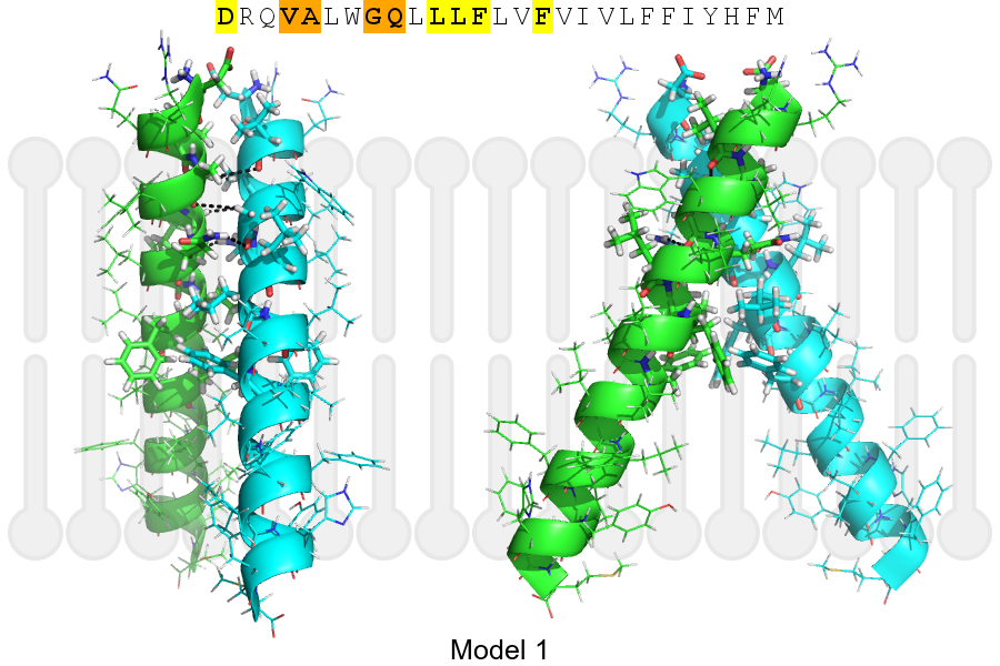
Click on the image to enlarge
Download model 1 structure: PDB, PyMOL session
List of hydrogen bonds:
| Donor | Acceptor | Distance |
|---|---|---|
| B, 224, ALA, HA | A, 223, VAL, O | 3.47Å |
| B, 227, GLY, HA1 | A, 224, ALA, O | 3.37Å |
| B, 228, GLN, HE21 | A, 227, GLY, O | 2.29Å |
| A, 224, ALA, HA | B, 223, VAL, O | 3.47Å |
| A, 227, GLY, HA1 | B, 224, ALA, O | 3.37Å |
| A, 228, GLN, HE21 | B, 227, GLY, O | 2.29Å |