

UniProt data
UniProt name: TGFR2_HUMAN (P37173)
Gene name: TGFBR2
Organism: Homo sapiens (Human).
Function: Transmembrane serine/threonine kinase forming with the TGF-beta type I serine/threonine kinase receptor, TGFBR1, the non- promiscuous receptor for the TGF-beta cytokines TGFB1, TGFB2 and TGFB3. Transduces the TGFB1, TGFB2 and TGFB3 signal from the cell surface to the cytoplasm and is thus regulating a plethora of physiological and pathological processes including cell cycle arrest in epithelial and hematopoietic cells, control of mesenchymal cell proliferation and differentiation, wound healing, extracellular matrix production, immunosuppression and carcinogenesis. The formation of the receptor complex composed of 2 TGFBR1 and 2 TGFBR2 molecules symmetrically bound to the cytokine dimer results in the phosphorylation and the activation of TGFRB1 by the constitutively active TGFBR2. Activated TGFBR1 phosphorylates SMAD2 which dissociates from the receptor and interacts with SMAD4. The SMAD2-SMAD4 complex is subsequently translocated to the nucleus where it modulates the transcription of the TGF-beta-regulated genes. This constitutes the canonical SMAD-dependent TGF-beta signaling cascade. Also involved in non- canonical, SMAD-independent TGF-beta signaling pathways. {ECO:0000269|PubMed:7774578}.
Subcellular location: Cell membrane {ECO:0000269|PubMed:1310899, ECO:0000269|PubMed:25893292}; Single-pass type I membrane protein {ECO:0000269|PubMed:1310899}. Membrane raft {ECO:0000269|PubMed:25893292}.
Transmembrane domains:
- 167-187, 21 AA, Helical. {ECO:0000255}.
Sequence: 567 AA
CATM prediction
The sequence of TGFR2_HUMAN returned 1 potential models
The positions at the dimer's interface are highlighted in yellow. The position at the crossing point is highlighted in orange.
| # | Sequence and interface | d | θ | Z' | ω' | Score |
|---|---|---|---|---|---|---|
| 170 180 1 + | + | + | |
(Å) | (°) | (Å) | (°) | ||
| Model 1 | IFQVTGISLLAALGVAISVIIIFYCYR | 7.4 | -48.0° | 0.5 | 47.2 | -9.7 |
| Prolines* | IFQVTGISLLPPLGVAISVIIIFYCYR |
* Note: this sequence contains Pro residues. CATM substituted them with Ala during the modeling. The positions are marked.
Click on the model number to visualize its details below.
MODEL 1 DETAILS
Precited structure:
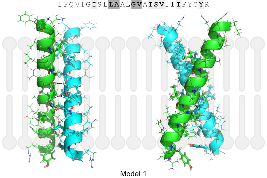
Click on the image to enlarge
Download model 1 structure: PDB, PyMOL session
List of hydrogen bonds:
| Donor | Acceptor | Distance |
|---|---|---|
| B, 177, GLY, HA1 | A, 174, ALA, O | 3.46Å |
| B, 178, VAL, HA | A, 177, GLY, O | 3.41Å |
| B, 181, SER, HG1 | A, 178, VAL, O | 2.35Å |
| A, 177, GLY, HA1 | B, 174, ALA, O | 3.46Å |
| A, 178, VAL, HA | B, 177, GLY, O | 3.41Å |
| A, 181, SER, HG1 | B, 178, VAL, O | 2.35Å |