UniProt data
UniProt name: EPHA1_HUMAN (P21709)
Gene name: EPHA1; Synonyms=EPH, EPHT, EPHT1
Organism: Homo sapiens (Human).
Function: Receptor tyrosine kinase which binds promiscuously membrane-bound ephrin-A family ligands residing on adjacent cells, leading to contact-dependent bidirectional signaling into neighboring cells. The signaling pathway downstream of the receptor is referred to as forward signaling while the signaling pathway downstream of the ephrin ligand is referred to as reverse signaling. Binds with a low affinity EFNA3 and EFNA4 and with a high affinity to EFNA1 which most probably constitutes its cognate/functional ligand. Upon activation by EFNA1 induces cell attachment to the extracellular matrix inhibiting cell spreading and motility through regulation of ILK and downstream RHOA and RAC. Plays also a role in angiogenesis and regulates cell proliferation. May play a role in apoptosis. {ECO:0000269|PubMed:17634955, ECO:0000269|PubMed:19118217, ECO:0000269|PubMed:20043122}.
Subcellular location: Cell membrane {ECO:0000269|PubMed:19118217}; Single-pass type I membrane protein {ECO:0000269|PubMed:19118217}.
Transmembrane domains:
- 548-568, 21 AA, Helical. {ECO:0000255}.
Sequence: 976 AA
CATM prediction
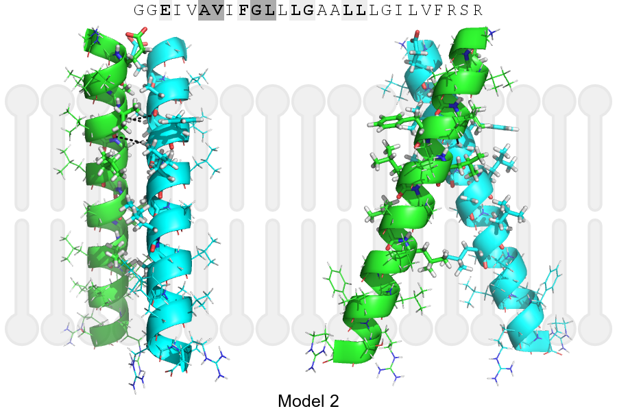
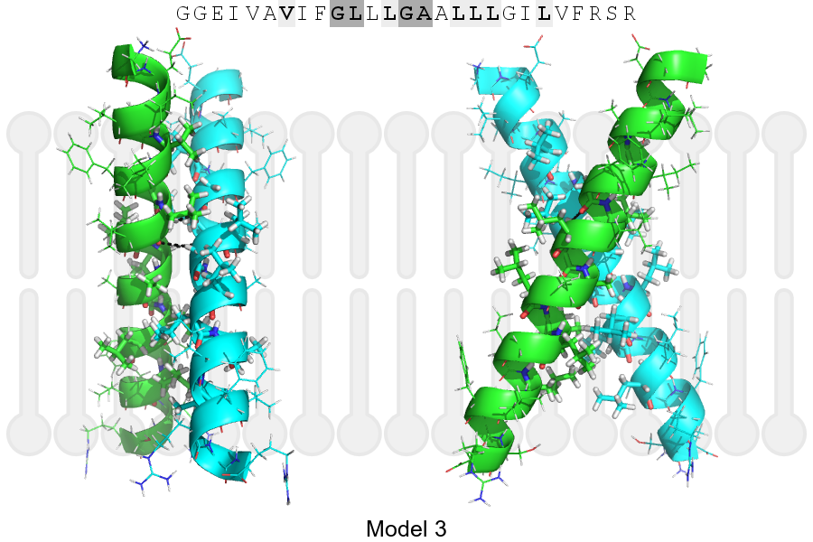
The sequence of EPHA1_HUMAN returned 3 potential models
The positions at the dimer's interface are highlighted in yellow. The position at the crossing point is highlighted in orange.
| # | Sequence and interface | d | θ | Z' | ω' | Score |
|---|---|---|---|---|---|---|
| 550 560 57 + | + | + | |
(Å) | (°) | (Å) | (°) | ||
| Model 1 | GGEIVAVIFGLLLGAALLLGILVFRSR | 7.2 | -44.0° | 1.8 | 59.2 | -29.8 |
| Model 2 | GGEIVAVIFGLLLGAALLLGILVFRSR | 7.2 | -44.0° | 4.9 | 44.0 | -9.2 |
| Model 3 | GGEIVAVIFGLLLGAALLLGILVFRSR | 6.4 | -55.0° | 2.4 | 47.6 | -7.0 |
Click on the model number to visualize its details below.
MODEL 1 DETAILS
Precited structure:
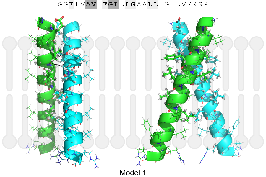
Click on the image to enlarge
Download model 1 structure: PDB, PyMOL session
List of hydrogen bonds:
| Donor | Acceptor | Distance |
|---|---|---|
| B, 551, VAL, HA | A, 550, ALA, O | 3.10Å |
| B, 554, GLY, HA1 | A, 551, VAL, O | 3.19Å |
| A, 551, VAL, HA | B, 550, ALA, O | 3.10Å |
| A, 554, GLY, HA1 | B, 551, VAL, O | 3.19Å |