

UniProt data
UniProt name: PECA1_HUMAN (P16284)
Gene name: PECAM1
Organism: Homo sapiens (Human).
Function: Induces susceptibility to atherosclerosis (By similarity). Cell adhesion molecule which is required for leukocyte transendothelial migration (TEM) under most inflammatory conditions. Tyr-690 plays a critical role in TEM and is required for efficient trafficking of PECAM1 to and from the lateral border recycling compartment (LBRC) and is also essential for the LBRC membrane to be targeted around migrating leukocytes. Prevents phagocyte ingestion of closely apposed viable cells by transmitting 'detachment' signals, and changes function on apoptosis, promoting tethering of dying cells to phagocytes (the encounter of a viable cell with a phagocyte via the homophilic interaction of PECAM1 on both cell surfaces leads to the viable cell's active repulsion from the phagocyte. During apoptosis, the inside-out signaling of PECAM1 is somehow disabled so that the apoptotic cell does not actively reject the phagocyte anymore. The lack of this repulsion signal together with the interaction of the eat-me signals and their respective receptors causes the attachment of the apoptotic cell to the phagocyte, thus triggering the process of engulfment). Isoform Delta15 is unable to protect against apoptosis. Modulates BDKRB2 activation. Regulates bradykinin- and hyperosmotic shock-induced ERK1/2 activation in human umbilical cord vein cells (HUVEC). {ECO:0000250, ECO:0000269|PubMed:12110892, ECO:0000269|PubMed:19342684}.
Subcellular location: Isoform Delta15: Cell junction {ECO:0000269|PubMed:18388311}. Note=Localizes to the lateral border recycling compartment (LBRC) and recycles from the LBRC to the junction in resting endothelial cells.
Transmembrane domains:
- 602-620, 19 AA, Helical. {ECO:0000255}.
Sequence: 738 AA
CATM prediction
The sequence of PECA1_HUMAN returned 1 potential models
The positions at the dimer's interface are highlighted in yellow. The position at the crossing point is highlighted in orange.
| # | Sequence and interface | d | θ | Z' | ω' | Score |
|---|---|---|---|---|---|---|
| 600 610 620 | + | + | |
(Å) | (°) | (Å) | (°) | ||
| Model 1 | WKKGLIAVVIIGVIIALLIIAAKCY | 7.2 | -50.9° | 1.2 | 44.4 | -14.6 |
Click on the model number to visualize its details below.
MODEL 1 DETAILS
Precited structure:
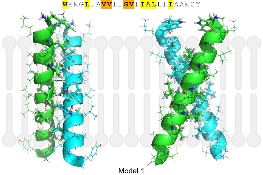
Click on the image to enlarge
Download model 1 structure: PDB, PyMOL session
List of hydrogen bonds:
| Donor | Acceptor | Distance |
|---|---|---|
| B, 610, GLY, HA1 | A, 607, VAL, O | 3.22Å |
| B, 611, VAL, HA | A, 610, GLY, O | 3.21Å |
| A, 610, GLY, HA1 | B, 607, VAL, O | 3.22Å |
| A, 611, VAL, HA | B, 610, GLY, O | 3.21Å |