

UniProt data
UniProt name: IL1R1_HUMAN (P14778)
Gene name: IL1R1; Synonyms=IL1R, IL1RA, IL1RT1
Organism: Homo sapiens (Human).
Function: Receptor for IL1A, IL1B and IL1RN. After binding to interleukin-1 associates with the coreceptor IL1RAP to form the high affinity interleukin-1 receptor complex which mediates interleukin-1-dependent activation of NF-kappa-B, MAPK and other pathways. Signaling involves the recruitment of adapter molecules such as TOLLIP, MYD88, and IRAK1 or IRAK2 via the respective TIR domains of the receptor/coreceptor subunits. Binds ligands with comparable affinity and binding of antagonist IL1RN prevents association with IL1RAP to form a signaling complex. {ECO:0000269|PubMed:10671496}.
Subcellular location: Membrane {ECO:0000269|PubMed:8142597}; Single-pass type I membrane protein {ECO:0000269|PubMed:8142597}. Cell membrane {ECO:0000305|PubMed:8142597}. Secreted {ECO:0000269|PubMed:8142597}.
Transmembrane domains:
- 337-356, 20 AA, Helical. {ECO:0000255}.
Sequence: 569 AA
CATM prediction
The sequence of IL1R1_HUMAN returned 1 potential models
The positions at the dimer's interface are highlighted in yellow. The position at the crossing point is highlighted in orange.
| # | Sequence and interface | d | θ | Z' | ω' | Score |
|---|---|---|---|---|---|---|
| 340 350 + | + | + |
(Å) | (°) | (Å) | (°) | ||
| Model 1 | FQKHMIGICVTLTVIIVCSVFIYKIF | 7.1 | -45.7° | 2.7 | 47.1 | -17.7 |
Click on the model number to visualize its details below.
MODEL 1 DETAILS
Precited structure:
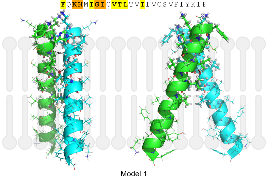
Click on the image to enlarge
Download model 1 structure: PDB, PyMOL session
List of hydrogen bonds:
| Donor | Acceptor | Distance |
|---|---|---|
| B, 340, GLY, HA1 | A, 337, HSE, O | 3.10Å |
| B, 341, ILE, HA | A, 344, THR, OG1 | 2.40Å |
| A, 340, GLY, HA1 | B, 337, HSE, O | 3.10Å |
| A, 341, ILE, HA | B, 344, THR, OG1 | 2.40Å |