

UniProt data
UniProt name: FGFR1_HUMAN (P11362)
Gene name: FGFR1; Synonyms=BFGFR, CEK, FGFBR, FLG, FLT2, HBGFR
Organism: Homo sapiens (Human).
Function: Tyrosine-protein kinase that acts as cell-surface receptor for fibroblast growth factors and plays an essential role in the regulation of embryonic development, cell proliferation, differentiation and migration. Required for normal mesoderm patterning and correct axial organization during embryonic development, normal skeletogenesis and normal development of the gonadotropin-releasing hormone (GnRH) neuronal system. Phosphorylates PLCG1, FRS2, GAB1 and SHB. Ligand binding leads to the activation of several signaling cascades. Activation of PLCG1 leads to the production of the cellular signaling molecules diacylglycerol and inositol 1,4,5-trisphosphate. Phosphorylation of FRS2 triggers recruitment of GRB2, GAB1, PIK3R1 and SOS1, and mediates activation of RAS, MAPK1/ERK2, MAPK3/ERK1 and the MAP kinase signaling pathway, as well as of the AKT1 signaling pathway. Promotes phosphorylation of SHC1, STAT1 and PTPN11/SHP2. In the nucleus, enhances RPS6KA1 and CREB1 activity and contributes to the regulation of transcription. FGFR1 signaling is down-regulated by IL17RD/SEF, and by FGFR1 ubiquitination, internalization and degradation. {ECO:0000250|UniProtKB:P16092, ECO:0000269|PubMed:10830168, ECO:0000269|PubMed:11353842, ECO:0000269|PubMed:12181353, ECO:0000269|PubMed:1379697, ECO:0000269|PubMed:1379698, ECO:0000269|PubMed:15117958, ECO:0000269|PubMed:16597617, ECO:0000269|PubMed:17311277, ECO:0000269|PubMed:17623664, ECO:0000269|PubMed:18480409, ECO:0000269|PubMed:19224897, ECO:0000269|PubMed:19261810, ECO:0000269|PubMed:19665973, ECO:0000269|PubMed:20133753, ECO:0000269|PubMed:20139426, ECO:0000269|PubMed:21765395, ECO:0000269|PubMed:8622701, ECO:0000269|PubMed:8663044}.
Subcellular location: Cell membrane; Single-pass type I membrane protein. Nucleus. Cytoplasm, cytosol. Cytoplasmic vesicle. Note=After ligand binding, both receptor and ligand are rapidly internalized. Can translocate to the nucleus after internalization, or by translocation from the endoplasmic reticulum or Golgi apparatus to the cytosol, and from there to the nucleus.
Transmembrane domains:
- 377-397, 21 AA, Helical. {ECO:0000255}.
Sequence: 822 AA
CATM prediction
The sequence of FGFR1_HUMAN returned 1 potential models
The positions at the dimer's interface are highlighted in yellow. The position at the crossing point is highlighted in orange.
| # | Sequence and interface | d | θ | Z' | ω' | Score |
|---|---|---|---|---|---|---|
| 380 390 4 + | + | + | |
(Å) | (°) | (Å) | (°) | ||
| Model 1 | YLEIIIYCTGAFLISCMVGSVIVYKMK | 7.3 | -50.9° | 3.3 | 48.1 | -25.6 |
Click on the model number to visualize its details below.
MODEL 1 DETAILS
Precited structure:
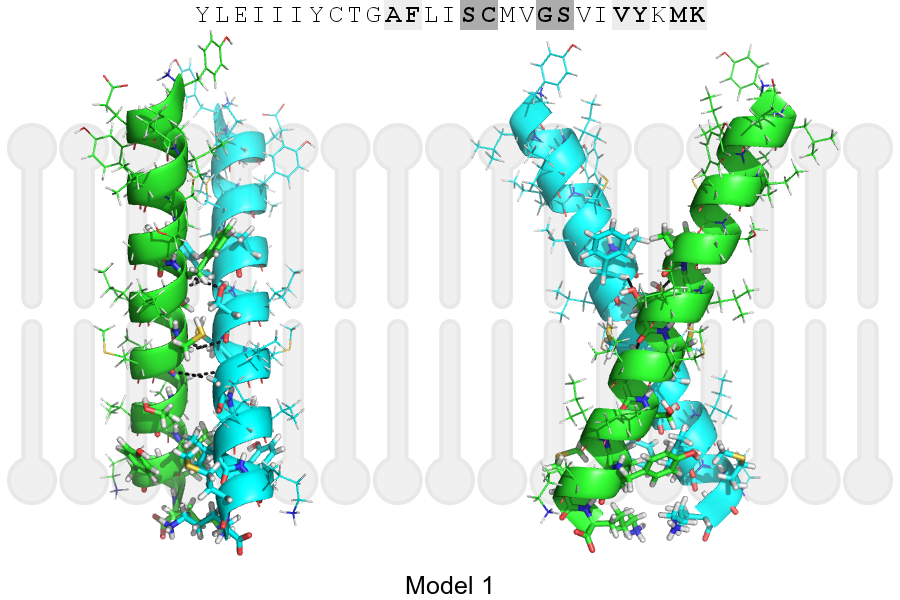
Click on the image to enlarge
Download model 1 structure: PDB, PyMOL session
List of hydrogen bonds:
| Donor | Acceptor | Distance |
|---|---|---|
| B, 385, PHE, HA | A, 388, SER, OG | 3.07Å |
| B, 389, CYS, HA | A, 388, SER, O | 3.14Å |
| B, 392, GLY, HA1 | A, 389, CYS, O | 3.28Å |
| A, 385, PHE, HA | B, 388, SER, OG | 3.09Å |
| A, 389, CYS, HA | B, 388, SER, O | 3.14Å |
| A, 392, GLY, HA1 | B, 389, CYS, O | 3.28Å |