UniProt data
UniProt name: 4F2_HUMAN (P08195)
Gene name: SLC3A2; Synonyms=MDU1
Organism: Homo sapiens (Human).
Function: Required for the function of light chain amino-acid transporters. Involved in sodium-independent, high-affinity transport of large neutral amino acids such as phenylalanine, tyrosine, leucine, arginine and tryptophan. Involved in guiding and targeting of LAT1 and LAT2 to the plasma membrane. When associated with SLC7A6 or SLC7A7 acts as an arginine/glutamine exchanger, following an antiport mechanism for amino acid transport, influencing arginine release in exchange for extracellular amino acids. Plays a role in nitric oxide synthesis in human umbilical vein endothelial cells (HUVECs) via transport of L-arginine. Required for normal and neoplastic cell growth. When associated with SLC7A5/LAT1, is also involved in the transport of L-DOPA across the blood-brain barrier, and that of thyroid hormones triiodothyronine (T3) and thyroxine (T4) across the cell membrane in tissues such as placenta. Involved in the uptake of methylmercury (MeHg) when administered as the L-cysteine or D,L-homocysteine complexes, and hence plays a role in metal ion homeostasis and toxicity. When associated with SLC7A5 or SLC7A8, involved in the cellular activity of small molecular weight nitrosothiols, via the stereoselective transport of L- nitrosocysteine (L-CNSO) across the transmembrane. Together with ICAM1, regulates the transport activity LAT2 in polarized intestinal cells, by generating and delivering intracellular signals. When associated with SLC7A5, plays an important role in transporting L-leucine from the circulating blood to the retina across the inner blood-retinal barrier. {ECO:0000269|PubMed:10903140, ECO:0000269|PubMed:11311135, ECO:0000269|PubMed:11389679, ECO:0000269|PubMed:11557028, ECO:0000269|PubMed:11564694, ECO:0000269|PubMed:11742812, ECO:0000269|PubMed:12117417, ECO:0000269|PubMed:12225859, ECO:0000269|PubMed:12716892, ECO:0000269|PubMed:14603368, ECO:0000269|PubMed:15769744, ECO:0000269|PubMed:15980244, ECO:0000269|PubMed:9751058, ECO:0000269|PubMed:9829974, ECO:0000269|PubMed:9878049}.
Subcellular location: Apical cell membrane; Single-pass type II membrane protein. Melanosome. Note=Identified by mass spectrometry in melanosome fractions from stage I to stage IV. Localized to the plasma membrane when associated with SLC7A5 or SLC7A8. Localized to the placental apical membrane. Located selectively at cell-cell adhesion sites (By similarity). Colocalized with SLC7A8/LAT2 at the basolateral membrane of kidney proximal tubules and small intestine epithelia. Expressed in both luminal and abluminal membranes of brain capillary endothelial cells (By similarity). {ECO:0000250}.
Transmembrane domains:
- 185-205, 21 AA, Helical; Signal-anchor for type II membrane protein. {ECO:0000255}.
Sequence: 630 AA
CATM prediction
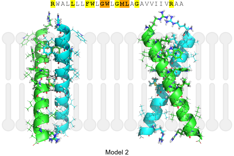
The sequence of 4F2_HUMAN returned 2 potential models
The positions at the dimer's interface are highlighted in yellow. The position at the crossing point is highlighted in orange.
| # | Sequence and interface | d | θ | Z' | ω' | Score |
|---|---|---|---|---|---|---|
| 190 200 + | + | + |
(Å) | (°) | (Å) | (°) | ||
| Model 1 | RWALLLLFWLGWLGMLAGAVVIIVRAA | 6.5 | -55.0° | 4.9 | 53.4 | -23.8 |
| Model 2 | RWALLLLFWLGWLGMLAGAVVIIVRAA | 6.9 | +49.1° | 2.8 | 88.0 | -19.0 |
| Prolines* | RWALLLLFWLGWLGMLAGAVVIIVRAP |
* Note: this sequence contains Pro residues. CATM substituted them with Ala during the modeling. The positions are marked.
Click on the model number to visualize its details below.
MODEL 1 DETAILS
Precited structure:
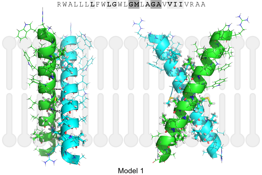
Click on the image to enlarge
Download model 1 structure: PDB, PyMOL session
List of hydrogen bonds:
| Donor | Acceptor | Distance |
|---|---|---|
| B, 195, GLY, HA1 | A, 192, GLY, O | 2.98Å |
| B, 196, MET, HA | A, 195, GLY, O | 2.39Å |
| B, 199, GLY, HA1 | A, 196, MET, O | 3.11Å |
| A, 195, GLY, HA1 | B, 192, GLY, O | 2.98Å |
| A, 196, MET, HA | B, 195, GLY, O | 2.39Å |
| A, 199, GLY, HA1 | B, 196, MET, O | 3.11Å |