UniProt data
UniProt name: A4_HUMAN (P05067)
Gene name: APP; Synonyms=A4, AD1
Organism: Homo sapiens (Human).
Function: N-APP binds TNFRSF21 triggering caspase activation and degeneration of both neuronal cell bodies (via caspase-3) and axons (via caspase-6).
Subcellular location: Membrane; Single-pass type I membrane protein. Membrane, clathrin-coated pit. Note=Cell surface protein that rapidly becomes internalized via clathrin-coated pits. During maturation, the immature APP (N-glycosylated in the endoplasmic reticulum) moves to the Golgi complex where complete maturation occurs (O-glycosylated and sulfated). After alpha-secretase cleavage, soluble APP is released into the extracellular space and the C-terminal is internalized to endosomes and lysosomes. Some APP accumulates in secretory transport vesicles leaving the late Golgi compartment and returns to the cell surface. Gamma-CTF(59) peptide is located to both the cytoplasm and nuclei of neurons. It can be translocated to the nucleus through association with APBB1 (Fe65). Beta-APP42 associates with FRPL1 at the cell surface and the complex is then rapidly internalized. APP sorts to the basolateral surface in epithelial cells. During neuronal differentiation, the Thr-743 phosphorylated form is located mainly in growth cones, moderately in neurites and sparingly in the cell body. Casein kinase phosphorylation can occur either at the cell surface or within a post-Golgi compartment. Associates with GPC1 in perinuclear compartments. Colocalizes with SORL1 in a vesicular pattern in cytoplasm and perinuclear regions.
Transmembrane domains:
- 700-723, 24 AA, Helical. {ECO:0000255}.
Sequence: 770 AA
CATM prediction
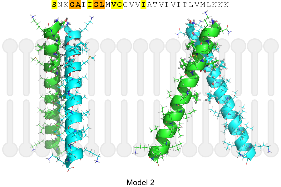
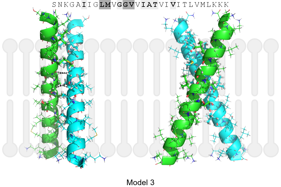
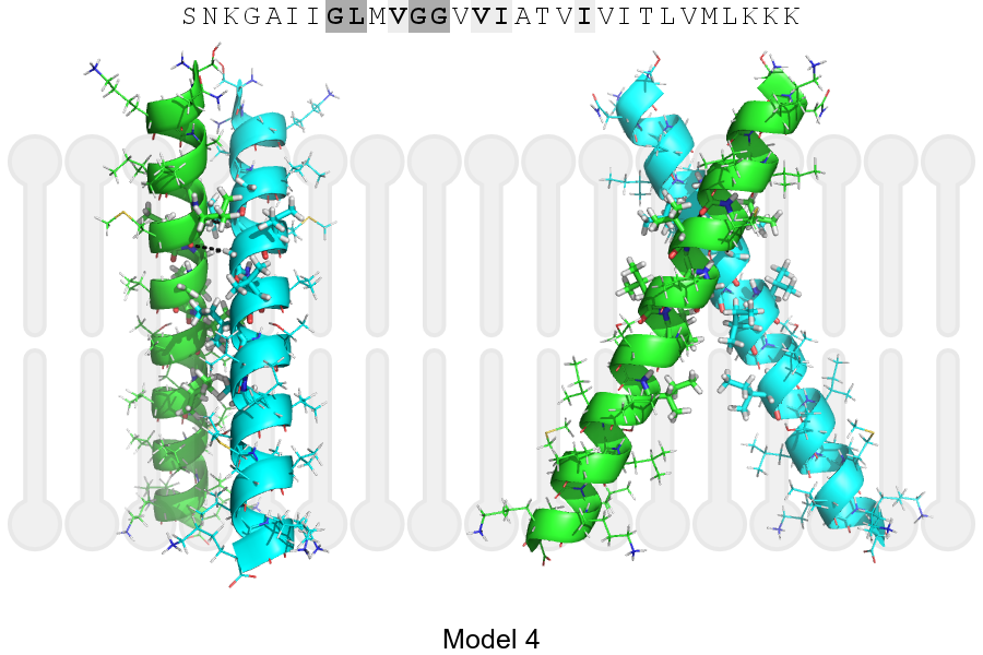
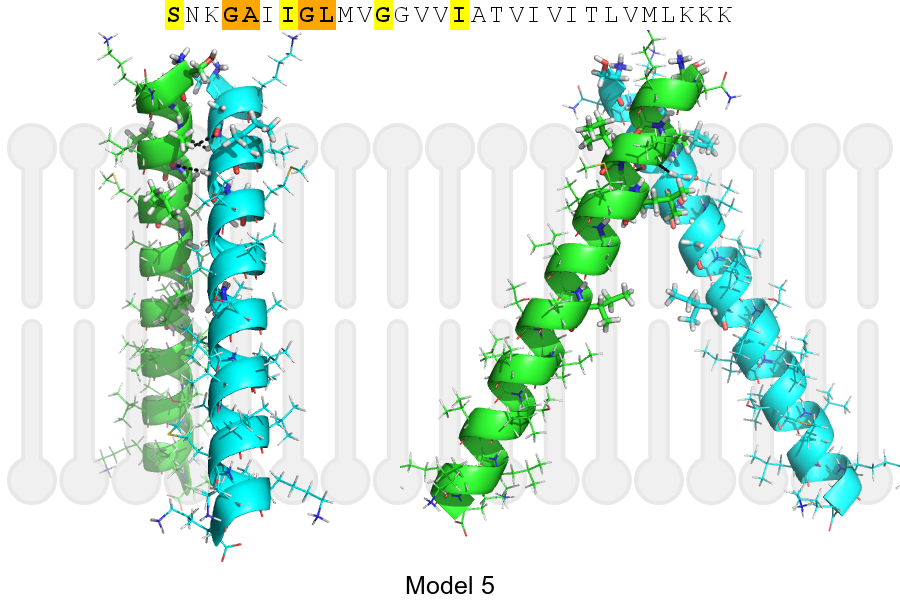
The sequence of A4_HUMAN returned 5 potential models
The positions at the dimer's interface are highlighted in yellow. The position at the crossing point is highlighted in orange.
| # | Sequence and interface | d | θ | Z' | ω' | Score |
|---|---|---|---|---|---|---|
| 700 710 720 | + | + | + |
(Å) | (°) | (Å) | (°) | ||
| Model 1 | SNKGAIIGLMVGGVVIATVIVITLVMLKKK | 6.7 | -28.2° | 6.0 | 47.0 | -30.8 |
| Model 2 | SNKGAIIGLMVGGVVIATVIVITLVMLKKK | 6.4 | -47.2° | 3.3 | 54.0 | -27.2 |
| Model 3 | SNKGAIIGLMVGGVVIATVIVITLVMLKKK | 7.4 | -48.1° | 0.4 | 47.0 | -23.3 |
| Model 4 | SNKGAIIGLMVGGVVIATVIVITLVMLKKK | 7.0 | -54.1° | 3.6 | 48.9 | -14.1 |
| Model 5 | SNKGAIIGLMVGGVVIATVIVITLVMLKKK | 6.4 | -55.1° | 5.9 | 46.8 | -10.4 |
Click on the model number to visualize its details below.
MODEL 1 DETAILS
Precited structure:
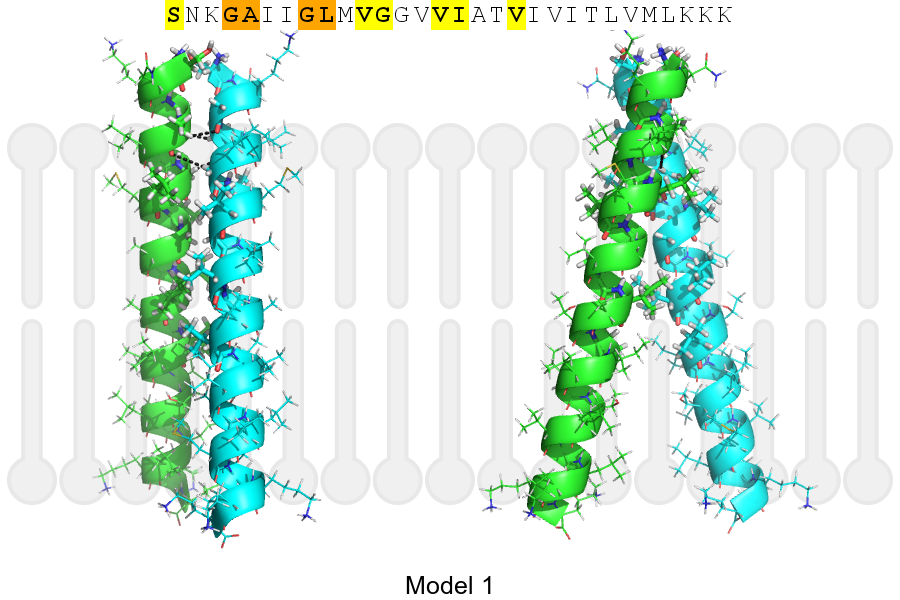
Click on the image to enlarge
Download model 1 structure: PDB, PyMOL session
List of hydrogen bonds:
| Donor | Acceptor | Distance |
|---|---|---|
| B, 701, ALA, HA | A, 700, GLY, O | 2.57Å |
| B, 704, GLY, HA1 | A, 701, ALA, O | 3.34Å |
| A, 701, ALA, HA | B, 700, GLY, O | 2.57Å |
| A, 704, GLY, HA1 | B, 701, ALA, O | 3.34Å |