UniProt data
UniProt name: GLPA_HUMAN (P02724)
Gene name: GYPA; Synonyms=GPA
Organism: Homo sapiens (Human).
Function: Glycophorin A is the major intrinsic membrane protein of the erythrocyte. The N-terminal glycosylated segment, which lies outside the erythrocyte membrane, has MN blood group receptors. Appears to be important for the function of SLC4A1 and is required for high activity of SLC4A1. May be involved in translocation of SLC4A1 to the plasma membrane. Is a receptor for influenza virus. Is a receptor for Plasmodium falciparum erythrocyte-binding antigen 175 (EBA-175); binding of EBA-175 is dependent on sialic acid residues of the O-linked glycans. Appears to be a receptor for Hepatitis A virus (HAV). {ECO:0000269|PubMed:10926825, ECO:0000269|PubMed:12813056, ECO:0000269|PubMed:14604989, ECO:0000269|PubMed:15331714, ECO:0000269|PubMed:19438409, ECO:0000269|PubMed:8009226}.
Subcellular location: Cell membrane {ECO:0000269|PubMed:11402026}; Single-pass type I membrane protein {ECO:0000269|PubMed:11402026}. Note=Appears to be colocalized with SLC4A1.
Transmembrane domains:
- 92-114, 23 AA, Helical.
Sequence: 150 AA
CATM prediction
The sequence of GLPA_HUMAN returned 3 potential models
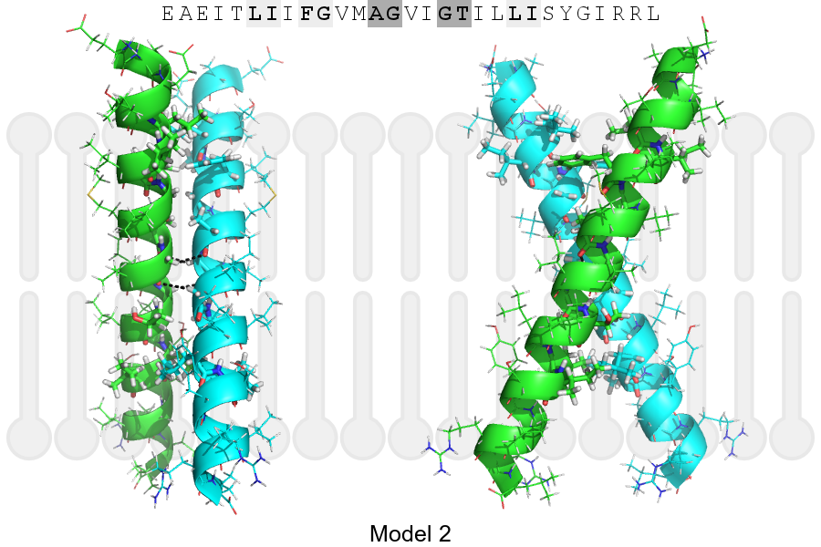
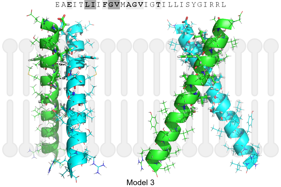
The positions at the dimer's interface are highlighted in yellow. The position at the crossing point is highlighted in orange.
| # | Sequence and interface | d | θ | Z' | ω' | Score |
|---|---|---|---|---|---|---|
| 90 100 110 | + | + | + |
(Å) | (°) | (Å) | (°) | ||
| Model 1 | EAEITLIIFGVMAGVIGTILLISYGIRRL | 6.7 | -49.1° | 4.5 | 48.8 | -18.1 |
| Model 2 | EAEITLIIFGVMAGVIGTILLISYGIRRL | 6.9 | -51.1° | 4.4 | 39.8 | -12.1 |
| Model 3 | EAEITLIIFGVMAGVIGTILLISYGIRRL | 7.0 | -54.8° | 1.5 | 44.6 | -6.7 |
| Prolines* | EPEITLIIFGVMAGVIGTILLISYGIRRL |
* Note: this sequence contains Pro residues. CATM substituted them with Ala during the modeling. The positions are marked.
Click on the model number to visualize its details below.
MODEL 1 DETAILS
Precited structure:
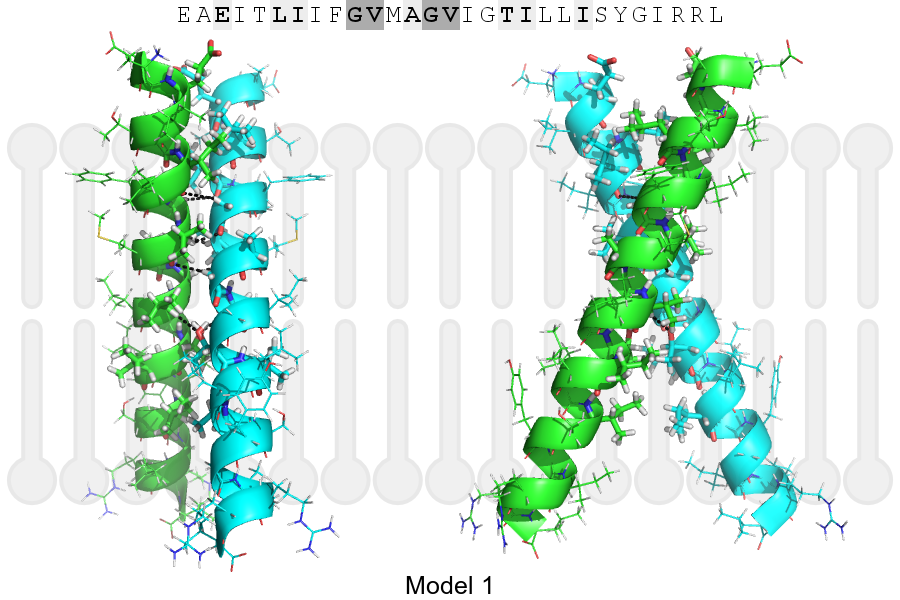
Click on the image to enlarge
Download model 1 structure: PDB, PyMOL session
List of hydrogen bonds:
| Donor | Acceptor | Distance |
|---|---|---|
| B, 98, GLY, HA1 | A, 95, ILE, O | 3.45Å |
| B, 99, VAL, HA | A, 98, GLY, O | 2.54Å |
| B, 102, GLY, HA1 | A, 99, VAL, O | 2.89Å |
| B, 103, VAL, HA | A, 106, THR, OG1 | 2.93Å |
| A, 98, GLY, HA1 | B, 95, ILE, O | 3.45Å |
| A, 99, VAL, HA | B, 98, GLY, O | 2.54Å |
| A, 102, GLY, HA1 | B, 99, VAL, O | 2.89Å |
| A, 103, VAL, HA | B, 106, THR, OG1 | 2.93Å |