

UniProt data
UniProt name: B3GA3_HUMAN (O94766)
Gene name: B3GAT3
Organism: Homo sapiens (Human).
Function: Glycosaminoglycans biosynthesis (PubMed:25893793). Involved in forming the linkage tetrasaccharide present in heparan sulfate and chondroitin sulfate. Transfers a glucuronic acid moiety from the uridine diphosphate-glucuronic acid (UDP-GlcUA) to the common linkage region trisaccharide Gal-beta-1,3-Gal-beta-1,4- Xyl covalently bound to a Ser residue at the glycosaminylglycan attachment site of proteoglycans. Can also play a role in the biosynthesis of l2/HNK-1 carbohydrate epitope on glycoproteins. Shows strict specificity for Gal-beta-1,3-Gal-beta-1,4-Xyl, exhibiting negligible incorporation into other galactoside substrates including Galbeta1-3Gal beta1-O-benzyl, Galbeta1- 4GlcNAc and Galbeta1-4Glc. Stimulates 2-phosphoxylose phosphatase activity of PXYLP1 in presence of uridine diphosphate-glucuronic acid (UDP-GlcUA) during completion of linkage region formation (PubMed:24425863). {ECO:0000269|PubMed:24425863, ECO:0000269|PubMed:25893793}.
Subcellular location: Golgi apparatus membrane {ECO:0000269|PubMed:21763480}; Single-pass type II membrane protein {ECO:0000269|PubMed:21763480}. Golgi apparatus, cis-Golgi network {ECO:0000269|PubMed:21763480, ECO:0000269|PubMed:25893793}.
Transmembrane domains:
- 8-28, 21 AA, Helical; Signal-anchor for type II membrane protein. {ECO:0000255}.
Sequence: 335 AA
CATM prediction
The sequence of B3GA3_HUMAN returned 1 potential models
The positions at the dimer's interface are highlighted in yellow. The position at the crossing point is highlighted in orange.
| # | Sequence and interface | d | θ | Z' | ω' | Score |
|---|---|---|---|---|---|---|
| 10 20 30 + | + | + | |
(Å) | (°) | (Å) | (°) | ||
| Model 1 | LKNVFLAYFLVSIAGLLYALVQLGQAC | 7.3 | -51.0° | 2.4 | 45.1 | -7.6 |
| Prolines* | LKNVFLAYFLVSIAGLLYALVQLGQPC |
* Note: this sequence contains Pro residues. CATM substituted them with Ala during the modeling. The positions are marked.
Click on the model number to visualize its details below.
MODEL 1 DETAILS
Precited structure:
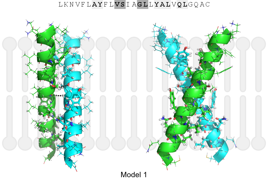
Click on the image to enlarge
Download model 1 structure: PDB, PyMOL session
List of hydrogen bonds:
| Donor | Acceptor | Distance |
|---|---|---|
| B, 16, SER, HA | A, 15, VAL, O | 3.46Å |
| B, 19, GLY, HA1 | A, 16, SER, O | 3.23Å |
| A, 16, SER, HA | B, 15, VAL, O | 3.46Å |
| A, 19, GLY, HA1 | B, 16, SER, O | 3.23Å |