UniProt data
UniProt name: BNI3L_HUMAN (O60238)
Gene name: BNIP3L; Synonyms=BNIP3A, BNIP3H, NIX
Organism: Homo sapiens (Human).
Function: Induces apoptosis. Interacts with viral and cellular anti-apoptosis proteins. Can overcome the suppressors BCL-2 and BCL-XL, although high levels of BCL-XL expression will inhibit apoptosis. Inhibits apoptosis induced by BNIP3. Involved in mitochondrial quality control via its interaction with SPATA18/MIEAP: in response to mitochondrial damage, participates in mitochondrial protein catabolic process (also named MALM) leading to the degradation of damaged proteins inside mitochondria. The physical interaction of SPATA18/MIEAP, BNIP3 and BNIP3L/NIX at the mitochondrial outer membrane regulates the opening of a pore in the mitochondrial double membrane in order to mediate the translocation of lysosomal proteins from the cytoplasm to the mitochondrial matrix. May function as a tumor suppressor. {ECO:0000269|PubMed:10381623, ECO:0000269|PubMed:21264228}.
Subcellular location: Nucleus envelope. Endoplasmic reticulum. Mitochondrion outer membrane. Membrane {ECO:0000305}; Single-pass membrane protein {ECO:0000305}. Note=Colocalizes with SPATA18 at the mitochondrion outer membrane.
Transmembrane domains:
- 188-208, 21 AA, Helical. {ECO:0000255}.
Sequence: 219 AA
CATM prediction
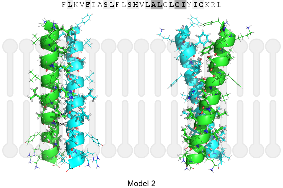
The sequence of BNI3L_HUMAN returned 2 potential models
The positions at the dimer's interface are highlighted in yellow. The position at the crossing point is highlighted in orange.
| # | Sequence and interface | d | θ | Z' | ω' | Score |
|---|---|---|---|---|---|---|
| 190 200 21 + | + | + | |
(Å) | (°) | (Å) | (°) | ||
| Model 1 | FLKVFIASLFLSHVLALGLGIYIGKRL | 6.3 | -40.9° | 0.0 | 59.0 | -43.1 |
| Model 2 | FLKVFIASLFLSHVLALGLGIYIGKRL | 7.0 | -28.9° | 2.5 | 60.1 | -21.9 |
| Prolines* | FLKVFIPSLFLSHVLALGLGIYIGKRL |
* Note: this sequence contains Pro residues. CATM substituted them with Ala during the modeling. The positions are marked.
Click on the model number to visualize its details below.
MODEL 1 DETAILS
Precited structure:
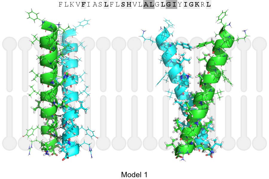
Click on the image to enlarge
Download model 1 structure: PDB, PyMOL session
List of hydrogen bonds:
| Donor | Acceptor | Distance |
|---|---|---|
| B, 201, LEU, HA | A, 200, ALA, O | 2.97Å |
| B, 204, GLY, HA1 | A, 201, LEU, O | 2.72Å |
| B, 205, ILE, HA | A, 204, GLY, O | 2.45Å |
| B, 208, GLY, HA1 | A, 205, ILE, O | 3.37Å |
| A, 201, LEU, HA | B, 200, ALA, O | 2.97Å |
| A, 204, GLY, HA1 | B, 201, LEU, O | 2.72Å |
| A, 205, ILE, HA | B, 204, GLY, O | 2.45Å |
| A, 208, GLY, HA1 | B, 205, ILE, O | 3.37Å |