UniProt data
UniProt name: TNF11_HUMAN (O14788)
Gene name: TNFSF11; Synonyms=OPGL, RANKL, TRANCE
Organism: Homo sapiens (Human).
Function: Cytokine that binds to TNFRSF11B/OPG and to TNFRSF11A/RANK. Osteoclast differentiation and activation factor. Augments the ability of dendritic cells to stimulate naive T-cell proliferation. May be an important regulator of interactions between T-cells and dendritic cells and may play a role in the regulation of the T-cell-dependent immune response. May also play an important role in enhanced bone-resorption in humoral hypercalcemia of malignancy (PubMed:22664871). Induces osteoclastogenesis by activating multiple signaling pathways in osteoclast precursor cells, chief among which is induction of long lasting oscillations in the intracellular concentration of Ca (2+) resulting in the activation of NFATC1, which translocates to the nucleus and induces osteoclast-specific gene transcription to allow differentiation of osteoclasts. During osteoclast differentiation, in a TMEM64 and ATP2A2-dependent manner induces activation of CREB1 and mitochondrial ROS generation necessary for proper osteoclast generation (By similarity). {ECO:0000250|UniProtKB:O35235, ECO:0000269|PubMed:22664871}.
Subcellular location: Tumor necrosis factor ligand superfamily member 11, soluble form: Secreted {ECO:0000250}.
Transmembrane domains:
- 48-68, 21 AA, Helical; Signal-anchor for type II membrane protein. {ECO:0000255}.
Sequence: 317 AA
CATM prediction
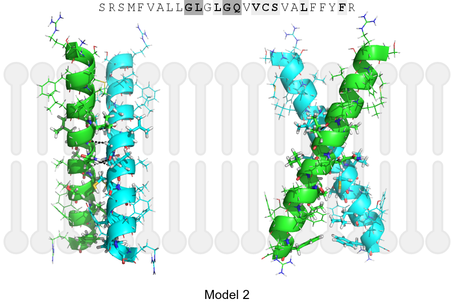
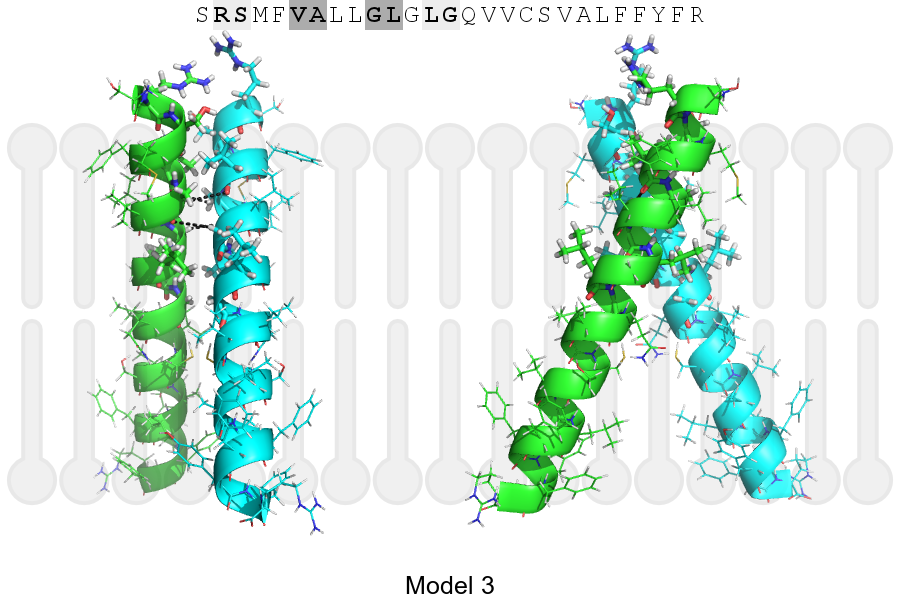
The sequence of TNF11_HUMAN returned 3 potential models
The positions at the dimer's interface are highlighted in yellow. The position at the crossing point is highlighted in orange.
| # | Sequence and interface | d | θ | Z' | ω' | Score |
|---|---|---|---|---|---|---|
| 50 60 70 + | + | + | |
(Å) | (°) | (Å) | (°) | ||
| Model 1 | SRSMFVALLGLGLGQVVCSVALFFYFR | 6.5 | -42.8° | 6.0 | 44.0 | -36.7 |
| Model 2 | SRSMFVALLGLGLGQVVCSVALFFYFR | 6.9 | -53.0° | 1.9 | 45.0 | -18.1 |
| Model 3 | SRSMFVALLGLGLGQVVCSVALFFYFR | 7.4 | -50.0° | 2.9 | 46.9 | -6.7 |
Click on the model number to visualize its details below.
MODEL 1 DETAILS
Precited structure:
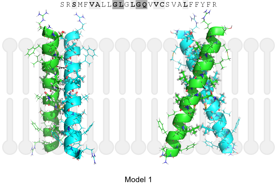
Click on the image to enlarge
Download model 1 structure: PDB, PyMOL session
List of hydrogen bonds:
| Donor | Acceptor | Distance |
|---|---|---|
| B, 51, ALA, HA | A, 50, VAL, O | 3.26Å |
| B, 54, GLY, HA1 | A, 51, ALA, O | 2.70Å |
| B, 55, LEU, HA | A, 54, GLY, O | 2.43Å |
| B, 58, GLY, HA1 | A, 55, LEU, O | 3.10Å |
| A, 51, ALA, HA | B, 50, VAL, O | 3.26Å |
| A, 54, GLY, HA1 | B, 51, ALA, O | 2.70Å |
| A, 55, LEU, HA | B, 54, GLY, O | 2.43Å |
| A, 58, GLY, HA1 | B, 55, LEU, O | 3.10Å |